. 2013 Aug 1;122(5):842-51.
doi: 10.1182/blood-2013-03-490219. Epub 2013 Jun 5.

Loss of endothelial protein C receptors links coagulation and inflammation to parasite sequestration in cerebral malaria in African children

Affiliations

Loss of endothelial protein C receptors links coagulation and inflammation to parasite sequestration in cerebral malaria in African children

Christopher A Moxon et al. Blood. .

Abstract

Cerebral malaria (CM) is a major cause of mortality in African children and the mechanisms underlying its development, namely how malaria-infected erythrocytes (IEs) cause disease and why the brain is preferentially affected, remain unclear. Brain microhemorrhages in CM suggest a clotting disorder, but whether this phenomenon is important in pathogenesis is debated. We hypothesized that localized cerebral microvascular thrombosis in CM is caused by a decreased expression of the anticoagulant and protective receptors thrombomodulin (TM) and endothelial protein C receptor (EPCR) and that low constitutive expression of these regulatory molecules in the brain make it particularly vulnerable. Autopsies from Malawian children with CM showed cerebral fibrin clots and loss of EPCR, colocalized with sequestered IEs. Using a novel assay to examine endothelial phenotype ex vivo using subcutaneous microvessels, we demonstrated that loss of EPCR and TM at sites of IE cytoadherence is detectible in nonfatal CM. In contrast, although clotting factor activation was seen in the blood of CM patients, this was compensated and did not disseminate. Because of the pleiotropic nature of EPCR and TM, these data implicate disruption of the endothelial protective properties at vulnerable sites and particularly in the brain, linking coagulation and inflammation with IE sequestration.


Figures
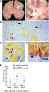
Figure 1
Figure 1
Microvascular thrombosis in association with IE sequestration in CM. (A) Coronal section of the brain from a child with fatal CM (right image detail of box in left) shows typical hemorrhagic lesions (arrow). (B) Low-power view (10× lens) shows fibrin deposition in microvessels in formalin-fixed paraffin-embedded tissue from CM case stained for fibrin using trichromic staining (fibrin stains red and red blood cells stain yellow) (C) Detail of box in 1B (40× lens) shows fibrin at the center of a ring hemorrhage (arrow). (D) Colocalization of fibrin and IE cells as indicated by malaria pigment (black dots, arrow). (E) Fibrin deposition was significantly higher in CM cases than in non-CM encephalopathic illness controls. The extent of the vessel lumen containing fibrin was scored as none, <50%, or ≥50 in 10 CM cases and 6 non-CM controls. Scoring was performed by the author and 2 blinded medical pathologists who each scored 50 vessels per case. Datapoints are a combination of the data from all 3 scorers and indicate the percentage of vessels scored at each level in individual cases and bars the mean percentage of vessels scored at each level for the CM or non-CM group as a whole. Micrographs were taken using a Leica DM1L microscope (Leica Microsystems) and a Micropublisher 3.3 RTV (QImaging) camera using Image ProPlus version 6.2 software (Media Cybernetics).
Figure 2
Figure 2
Loss of EPCR in the brain in CM and of TM in subcutaneous tissue. (A) Low-power (10× lens) view of postmortem brain samples shows vessels with moderate-to-strong staining for EPCR (immunoperoxidase method, arrows) in a non-CM control brain sample (left) and absence of EPCR staining in a vessel containing many sequestered IEs (arrow) in a CM case (right). (B) Association between IE sequestration, loss of EPCR, and fibrin deposition; upper image is a detail (40× lens) of the box in the bottom right of 2A and the lower image shows fibrin deposition (trichromic staining) in a consecutive tissue section. (C) EPCR staining is significantly reduced in CM compared with non-CM encephalopathic controls. The intensity of staining for EPCR was scored as none, weak, moderate (mod), or strong in 5 CM cases and 5 controls compared with reference images by the author and 2 independent pathologists. Datapoints are a combination of the data from all 3 scorers and indicate the percentage of vessels scored at each level in individual cases and bars the mean percentage of vessels scored at each level for the CM or non-CM group as a whole. (D) Subcutaneous tissue section in a fatal CM case. The red arrow indicates a vessel with high IE sequestration and minimal TM staining and the black arrow indicates a vessel with minimal IE sequestration and strong TM staining.
Figure 3
Figure 3
Endothelial activation and decreased EPCR and TM ex vivo in biopsies from children with CM. (A) Immunofluorescence-labeled needle biopsies samples of subcutaneous tissue from healthy children and children with CM. Nuclei appear blue (DAPI) and endothelial receptors green (Alexafluor 488). Micrographs (40× lens) show microvessels from healthy children as indicated by morphology, elliptical endothelial nuclei, and bright staining for CD31, ICAM-1, TM, and EPCR. Vessels from a CM case show IE sequestration (arrows) associated with low TM and EPCR staining. Needle biopsy samples were digested, labeled, and then analyzed by flow cytometer to examine the receptor expression of endothelial cells from microvessels in the sample. (B) Flow cytometry gating strategy for samples to distinguish endothelial cells as single, live, CD31+CD45− cells. (C) Histograms for 3 different endothelial receptors: ICAM-1, TM, and EPCR; representative plots from a single case for CM (blue), healthy children (HC; red), and antibody isotype control (IC, gray). ICAM-1 and TM staining show low overlap with the isotype control and receptor expression was determined by mean fluorescence intensity (MFI), whereas EPCR staining overlapped considerably with the isotype and expression was determined by percentage positive events. (D) Scatterplots for the endothelial expression levels of ICAM-1, TM, and EPCR as determined by flow cytometry in 17 CM cases and 20 HCs. (E) Scatterplots for levels of soluble ICAM-1, soluble TM, and soluble EPCR as determined by enzyme-linked immunosorbent assay in plasma samples in HC, CM, and aparasitemic febrile controls (FC) who were noncomatose patients that had a lumbar puncture taken because of clinical suspicion of meningitis. (F) Scatterplots for levels of soluble ICAM-1, soluble TM, and soluble EPCR in paired CSF samples in FC and CM; CSF samples were not taken from healthy children. Horizontal lines indicate geometric means and bars 95% CIs. Significance determined by one-way analysis of variance with the Tukey honestly significant difference test to adjust for multiple comparisons in E. * P < .05, ** P < .01, *** P < .001. Fluorescence micrographs were taken using a Leica DM1L microscope and a Leica DFC300FX camera using Leica Application Suite version 2.6.0 software.
Figure 4
Figure 4
Compensated activation of coagulation in CM. (A) Plasma TAT levels taken on admission in children with (right to left) retinopathy-positive CM (Ret Pos CM, n = 67), retinopathy-negative CM (Ret Neg CM, n = 19), nonmalarial coma (n = 11), uncomplicated malaria (n = 30), mild nonmalarial febrile illness (n = 30), and healthy controls (n = 19). (B) Plasma TAT levels taken on admission in patients with retinopathy-positive CM grouped according to outcome: those who eventually died (fatal [n = 16] and those who survived [nonfatal, n = 51]). (C) Prothrombin time and aPPT (D) in children with (from right to left) Ret Pos CM (n = 69), Ret Neg CM (n = 23), nonmalarial coma (n = 11), uncomplicated malaria (n = 21), mild nonmalarial febrile illness (n = 24), and healthy controls (n = 30). (E) aPC levels in plasma taken on admission in children with (from right to left): Ret Pos CM (n = 92), Ret Neg CM (n = 22), nonmalarial coma (n = 6), uncomplicated malaria (n = 24), mild nonmalarial febrile illness (n = 25), and healthy controls (n = 21). (F) Plasma aPC levels in retinopathy-positive children who eventually died (fatal, n = 16) and those who survived (nonfatal, n = 76). (G) Prothrombin fragment (F1+2)/aPC ratio in the same patients as 4E. (H) Prothrombin fragment levels in fatal and nonfatal retinopathy-positive CM. Horizontal lines indicate geometric means. Significance determined by ANOVA with the Tukey honestly significant difference test test on log-transformed data to adjust for multiple comparison. *P < .05, **P < .01, ***P < .001.
Figure 5
Figure 5
Sequestration-induced loss of protein C receptors links coagulation, inflammation, and endothelial permeability. PAR1 activation by thrombin acts as a molecular switch, inhibiting or promoting inflammation and leakage, depending on whether there is a modifying signal from the protein C pathway. Thrombin is produced by the interaction between circulating activated factor VII (VIIa) in the plasma and tissue factor on monocytes and from endothelial tissue factor induced by IE (step 1 in both A and B). (A) Thrombin/PAR1 signaling when the protein C system is intact; 2) thrombin initiates the TM/EPCR-facilitated activation of protein C, which inhibits thrombin production upstream; 3) aPC modifies the effect of PAR1 through EPCR; 4) aPC/EPCR-modified PAR1 signaling decreases endothelial permeability via S1P1 signaling (not shown) and production of S1P, which leads to enhancement of tight junctions; 5) in the presence of EPCR has pleotropic antiinflammatory and endothelial protective properties, including downregulation of Nuclear Factor κ-B (NFKB) and increased Angiopoetin-1 (Ang1) production. Ang1 decreases Weibel Palade body (WPB) exocytosis by occupancy of Tie2. (B) Thrombin/PAR1 signaling in a vessel with high level of sequestered malaria-IEs when there is complete loss of protein C receptors, such as in microvessels in the brain, and therefore no modification of PAR1 signaling. 2) IE sequestration is associated with loss of TM and EPCR; protein C is therefore not activated; 3) thrombin signals through PAR1 without modification by EPCR signaling; 4) unmodified PAR1 signaling inhibits S1P release with resultant loss of tight junctions, loss of endothelial barrier function, and localized vascular leak; and 5) thrombin signaling in the absence of modification by aPC/EPCR has strong proinflammatory effects, including upregulation of NFKB with increased tumor necrosis factor (TNF) and interleukin (IL)-6 production and reduction of Ang1 production, leading to increased Weibel Palade body (WPB) exocytosis with production of Von Willebrand Factor (vWF) and Ang2. Ang2 further increases WPB exocytosis by occupancy of Tie2 and also contributes to loss of endothelial barrier integrity and leak. 6) Thrombin and inflammatory cytokines cause activation of platelets, leading to the production of platelet microparticles. 7) In the absence of inhibition from aPC, thrombin triggers the production of fibrin from fibrinogen and fibrin and activated platelets coalesce to form thrombi. 8) Activated platelets adhere to vWF strings. Both thrombi and these platelet-vWF complexes impair cerebral circulation. Solid black arrows indicate stimulation of a pathway and dotted red lines indicate inhibition.

Comment in

Similar articles

Cited by

References

    1. Murray CJ, Rosenfeld LC, Lim SS, et al. Global malaria mortality between 1980 and 2010: a systematic analysis. Lancet. 2012;379(9814):413–431. - PubMed
    1. Taylor TE, Fu WJ, Carr RA, et al. Differentiating the pathologies of cerebral malaria by postmortem parasite counts. Nat Med. 2004;10(2):143–145. - PubMed
    1. Birbeck GL, Molyneux ME, Kaplan PW, et al. Blantyre Malaria Project Epilepsy Study (BMPES) of neurological outcomes in retinopathy-positive paediatric cerebral malaria survivors: a prospective cohort study. Lancet Neurol. 2010;9(12):1173–1181. - PMC - PubMed
    1. Seydel KB, Milner DA, Jr, Kamiza SB, Molyneux ME, Taylor TE. The distribution and intensity of parasite sequestration in comatose Malawian children. J Infect Dis. 2006;194(2):208–5. - PMC - PubMed
    1. Grau GE, Craig AG. Cerebral malaria pathogenesis: revisiting parasite and host contributions. Future Microbiol. 2012;7(2):291–302. - PubMed

Publication types

MeSH terms