. 2013 Sep;110(5):1097-106.
doi: 10.1152/jn.00114.2013. Epub 2013 Jun 5.

Vasoactive intestinal peptide produces long-lasting changes in neural activity in the suprachiasmatic nucleus

Affiliations

Vasoactive intestinal peptide produces long-lasting changes in neural activity in the suprachiasmatic nucleus

Takashi Kudo et al. J Neurophysiol. 2013 Sep.

Abstract

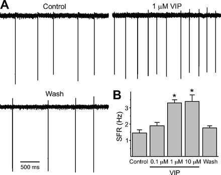
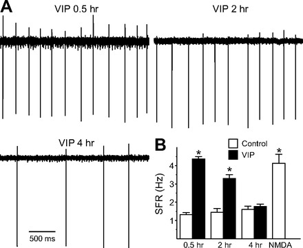
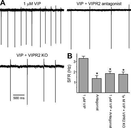
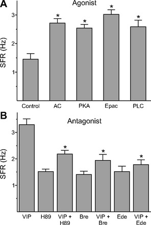
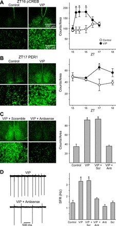
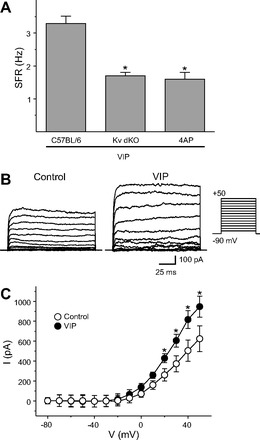
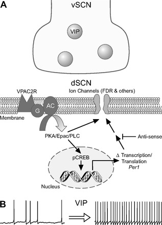
The neuropeptide vasoactive intestinal peptide (VIP) is expressed at high levels in the neurons of the suprachiasmatic nucleus (SCN). While VIP is known to be important to the input and output pathways from the SCN, the physiological effects of VIP on electrical activity of SCN neurons are not well known. Here the impact of VIP on firing rate of SCN neurons was investigated in mouse slice cultures recorded during the night. The application of VIP produced an increase in electrical activity in SCN slices that lasted several hours after treatment. This is a novel mechanism by which this peptide can produce long-term changes in central nervous system physiology. The increase in action potential frequency was blocked by a VIP receptor antagonist and lost in a VIP receptor knockout mouse. In addition, inhibitors of both the Epac family of cAMP binding proteins and cAMP-dependent protein kinase (PKA) blocked the induction by VIP. The persistent increase in spike rate following VIP application was not seen in SCN neurons from mice deficient in Kv3 channel proteins and was dependent on the clock protein PER1. These findings suggest that VIP regulates the long-term firing rate of SCN neurons through a VIPR2-mediated increase in the cAMP pathway and implicate the fast delayed rectifier (FDR) potassium currents as one of the targets of this regulation.

Keywords: VIP; circadian system; fast delayed rectifier; potassium; suprachiasmatic nucleus.


Figures

Fig. 1.
Fig. 1.
Application of vasoactive intestinal peptide (VIP) increased the firing rate of dorsal suprachiasmatic nucleus (dSCN) neurons during the night. A: representative examples illustrating the VIP-induced increase in firing rate in a dSCN neuron. B: average firing rate [spontaneous firing rate (SFR)] for each group (+SE). *Significant difference (P < 0.05) compared with controls analyzed by 1-way ANOVA followed by Dunn's method for multiple comparisons. The washout data were collected 4–6 h after drug application. For each group, n = 29 or 30 neurons. Throughout this study, sample sizes are reported as the number of neurons, with the data from each group coming from between 3 and 5 mice.
Fig. 2.
Fig. 2.
VIP produced long-lasting changes in firing rate in dSCN neurons during the night. A: representative examples illustrating the time course of VIP (1 μM)-evoked increase in firing rate. B: average firing rate for each group (+SE). *Significant difference (P < 0.05) analyzed by 2-way ANOVA followed by Holm-Sidak method for multiple comparisons (vs. control). For each group, n = 21–30 neurons.
Fig. 3.
Fig. 3.
VIP-induced increase in spike frequency is mediated by VIPR2. A: representative examples showing that VIP does not increase firing in the presence of the VIPR2 antagonist (MyrH-SDAVFTDNYTKLRKQMAVKKYLNSI-K-K-G-G-T, 1 μM) as well as in VIPR2 knockout (KO) mice. B: average firing rate for each group (+SE). *Significant difference (P < 0.05) analyzed by 1-way ANOVA followed by Holm-Sidak method for multiple comparisons (vs. 1 μM VIP). For each group, n = 30.
Fig. 4.
Fig. 4.
VIP-induced increase in spike frequency was mediated by both adenylyl cyclase (AC) and phospholipase C (PLC) pathways. A: average firing rate for the activators (+SE). *Significant difference (P < 0.05) analyzed by 1-way ANOVA followed by Dunn's method for multiple comparisons (vs. control). We evaluated several agents: AC activator forskolin (n = 14); cAMP-dependent protein kinase (PKA) activator N6-benzoyladenosine-3′,5′-cyclic monophosphate (6-Bnz-cAMP, n = 30); Epac activator cAMP receptor 8-(4-chlorophenylthio)-2′-O-methyl-cAMPS (n = 29); PLC activator m-3M3FBS (n = 30). B: average firing rate (+SE) for groups treated with the inhibitors alone and the inhibitors + VIP (1 μM). *Significant difference (P < 0.05) analyzed by 1-way ANOVA followed by Dunn's method for multiple comparisons (vs. VIP). We evaluated several agents: PKA inhibitor H89 (10 μM, n = 23); Epac inhibitor brefeldin A (Bre, 100 μM, n = 28); PLC inhibitor edelfosine (Ede, 10 μM, n = 24).
Fig. 5.
Fig. 5.
Persistent VIP-induced increases in electrical activity are dependent on PER1. A: photomicrographs illustrate that the application of VIP (1 μM) increases phospho-cAMP response element-binding protein (p-CREB) as measured by immunohistochemistry (IHC). Bar graphs show p-CREB-positive cell counts for control and VIP-treated groups (±SE). *Significant difference (P < 0.05) analyzed by 1-way ANOVA followed by Dunn's method for multiple comparisons (vs. control). ZT, zeitgeber time. B: photomicrographs illustrate that application of VIP (1 μM) increases PER1 protein as measured by IHC. Bar graphs show PER1-positive cell counts for control and VIP-treated groups (±SE). *Significant difference (P < 0.05) analyzed by 1-way ANOVA followed by Dunn's method for multiple comparisons (vs. control). C: photomicrographs illustrate that antisense against Per1 blocked the VIP induction of PER1. Bar graphs show PER1-positive cell counts for control and VIP-treated groups (+SE) exposed to antisense or scrambled message. Anti, group treated with antisense; Scr, group treated with scrambled message. D: representative examples showing that the application of VIP (1 μM) does not cause persistent increase in neural activity in the presence of antisense against Per1. Bar graphs show average firing rate for each group (+SE). *Significant difference (P < 0.05) analyzed by 1-way ANOVA followed by Dunn's method for multiple comparisons.
Fig. 6.
Fig. 6.
VIP-induced increase spike frequency was mediated by fast delayed rectifier (FDR) current. A: average firing rate for each group (+SE). *Significant difference (P < 0.05) analyzed by 1-way ANOVA followed by Dunn's method for multiple comparisons (vs. VIP). For each, VIP was applied at 1 μM and 4-aminopyridine (4-AP) at 0.5 mM. For each group, n = 30. B: representative examples showing that application of VIP (1 μM) increases the magnitude of FDR currents in dSCN. C: current (I)-voltage (V) relationship of FDR currents in SCN neurons. Data are shown as means ± SE. *Significant difference (P < 0.05) analyzed by 2-way repeated-measures ANOVA followed by Holm-Sidak method for multiple comparisons (vs. control). For each group, n = 11 or 12. FDR currents were isolated by subtraction (baseline, 0.5 mM 4-AP) using a voltage-step protocol with a prepulse potential of −90 mV and test pulse potentials (see materials and methods). Holding potential was −70 mV.
Fig. 7.
Fig. 7.
An illustration of our model of VIP regulation of firing rate in the dSCN. A: VIP is held in dense core synaptic vesicles in a subset of neurons in the ventral SCN (vSCN) region. Upon release, VIP activates the VPAC2 (VIPR2) receptors and activates a network of signaling pathways. These pathways produce short-term changes in firing rate presumably through posttranslational modification of intrinsic ion channels. In addition, these signaling pathways phosphorylate CREB and increase the transcription and translation of the clock gene Period1 (Per1). The increase in PER1 regulates the FDR current among other intrinsic ion channels to increase the firing rate of dSCN neurons. B: the net result of VIP application is a long-lasting (2–4 h) increase in the firing rate of dSCN neurons. It remains to be seen whether the ability of VIP to modulate ongoing electrical activity over the course of hours is restricted to the SCN or whether this is a common mechanism by which this peptide regulates nervous system function.

References

    1. Akiyama M, Kouzu Y, Takahashi S, Wakamatsu H, Moriya T, Maetani M, Watanabe S, Tei H, Sakaki Y, Shibata S. Inhibition of light- or glutamate-induced mPer1 expression represses the phase shifts into the mouse circadian locomotor and suprachiasmatic firing rhythms. J Neurosci 19: 1115–1121, 1999 - PMC - PubMed
    1. An S, Irwin RP, Allen CN, Tsai C, Herzog ED. Vasoactive intestinal polypeptide requires parallel changes in adenylate cyclase and phospholipase C to entrain circadian rhythms to a predictable phase. J Neurophysiol 105: 2289–2296, 2011 - PMC - PubMed
    1. An S, Tsai C, Ronecker J, Bayly A, Herzog ED. Spatiotemporal distribution of vasoactive intestinal polypeptide receptor 2 in mouse suprachiasmatic nucleus. J Comp Neurol 520: 2730–2741, 2012 - PMC - PubMed
    1. Aton SJ, Colwell CS, Harmar AJ, Waschek J, Herzog ED. Vasoactive intestinal polypeptide mediates circadian rhythmicity and synchrony in mammalian clock neurons. Nat Neurosci 8: 476–483, 2005 - PMC - PubMed
    1. Baranauskas G. Ionic channel function in action potential generation: current perspective. Mol Neurobiol 35: 129–150, 2007 - PubMed

MeSH terms

Substances

LinkOut - more resources