

Multiple cervical ganglioneuroma: A case report and review of the literature
- PMID: 23741250
- PMCID: PMC3673638
- DOI: 10.3892/ol.2012.767
Multiple cervical ganglioneuroma: A case report and review of the literature
Abstract
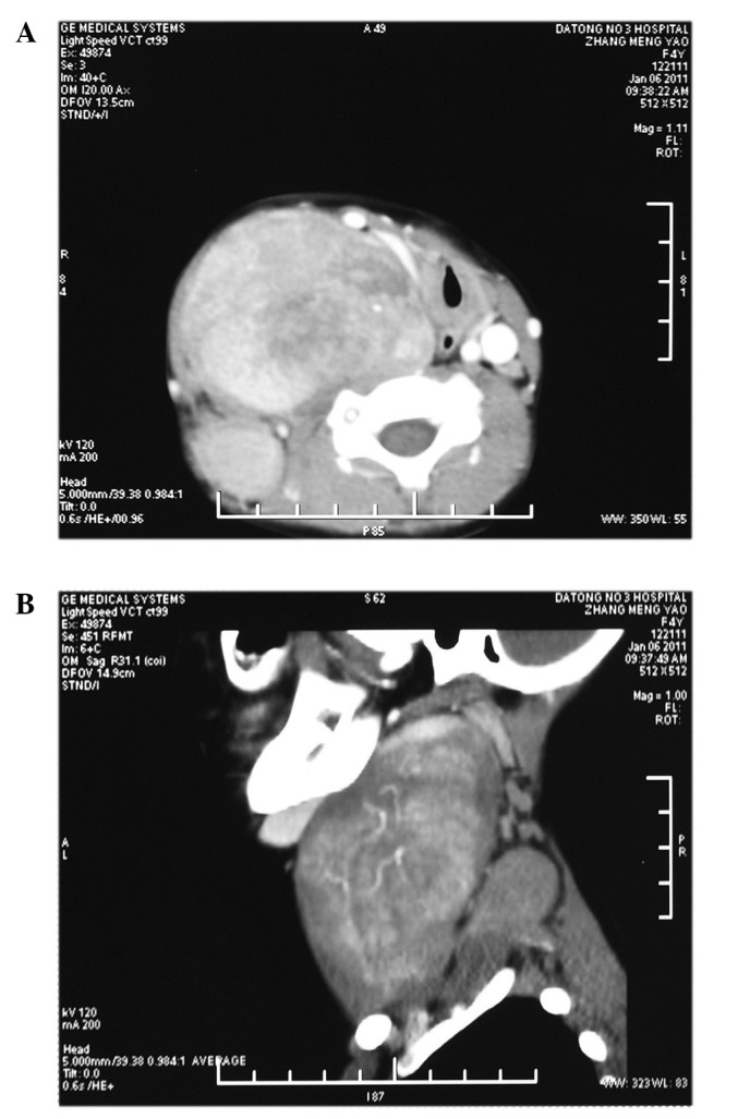
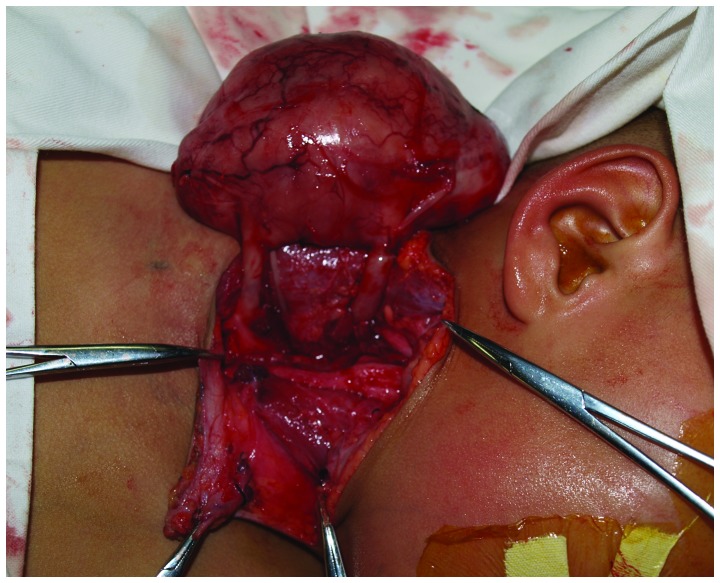
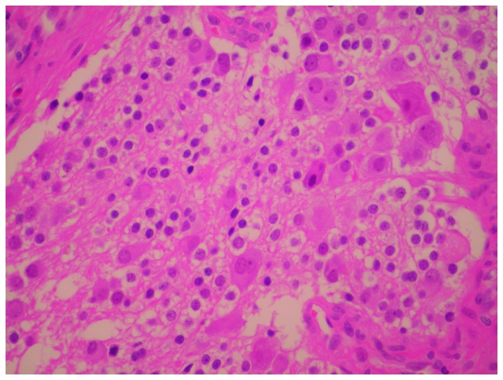
Ganglioneuromas (GNs) arising from neural crest sympathogonia are rare benign neurogenic tumors. The most commonly affected sites are the posterior mediastinum, the retroperitoneum and the adrenal gland. GNs often present as a solitary, painless and slow-growing mass, and multiple occurrences in the cervical region are extremely rare. Here, we report a case of massive multiple cervical GN in a 4-year-old girl, and review cases of cervical GN that have been reported in the past 10 years. The results demonstrated that cervical GN, compared to other sites, is seldom secretory. The signs and symptoms of cervical GN are unspecific; the ultimate diagnosis of GN depends on pathological examination. Fine-needle aspiration biopsy has limited value in diagnosis. Surgical excision is the treatment of choice and the prognosis is excellent even in cases where complete excision cannot be achieved. Furthermore, GNs should be considered in patients with multiple masses in the neck.
Keywords: cervical; children; ganglioneuroma; neurogenic tumor.
Figures
References
-
- Shotton JC, Milton CM, Allen JP. Multiple ganglioneuroma of the neck. J Laryngol Otol. 1992;106:277–278. - PubMed
-
- Miyakoshi N, Hongo M, Kasukawa Y, Misawa A, Shimada Y. Bilateral and symmetric C1–C2 dumbbell ganglioneuromas associated with neurofibromatosis type 1 causing severe spinal cord compression. Spine J. 2010;10:e11–e15. - PubMed
-
- Bacci C, Sestini R, Ammannati F, Bianchini E, Palladino T, Carella M, Melchionda S, Zelante L, Papi L. Multiple spinal ganglioneuromas in a patient harboring a pathogenic NF1 mutation. Clin Genet. 2010;77:293–297. - PubMed
-
- Zebing Z, Jianwei S, Yan C, Yan G. Clinicopathological characteristics of neck ganglioneuroma. Oral Med Pathol. 2008;12:131–134.
-
- Patterson AR, Barker CS, Loukota RA, Spencer J. Ganglioneuroma of the mandible resulting from metastasis of neuroblastoma. Int J Oral Maxillofac Surg. 2009;38:196–198. - PubMed
LinkOut - more resources
Full Text Sources
Miscellaneous