

Cholecalciferol (vitamin D₃) improves myelination and recovery after nerve injury
- PMID: 23741446
- PMCID: PMC3669361
- DOI: 10.1371/journal.pone.0065034
Cholecalciferol (vitamin D₃) improves myelination and recovery after nerve injury
Abstract
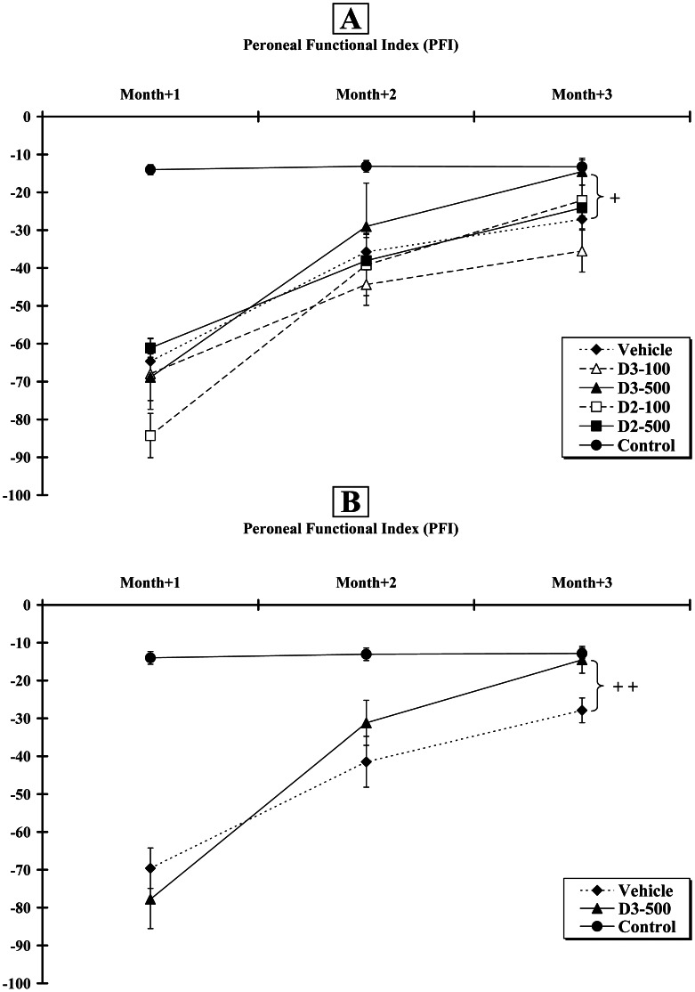
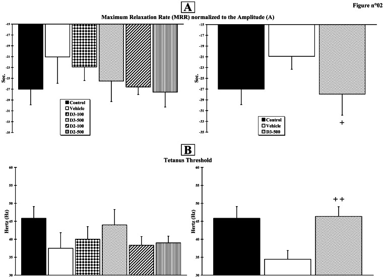
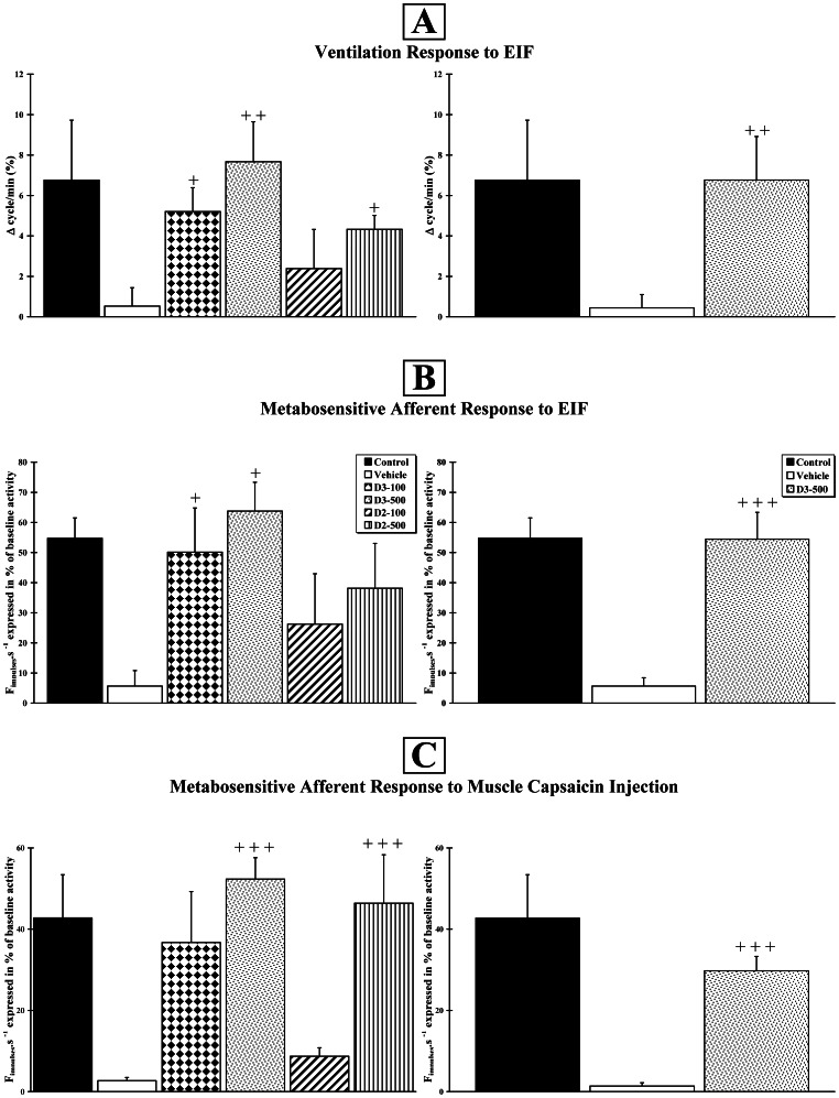
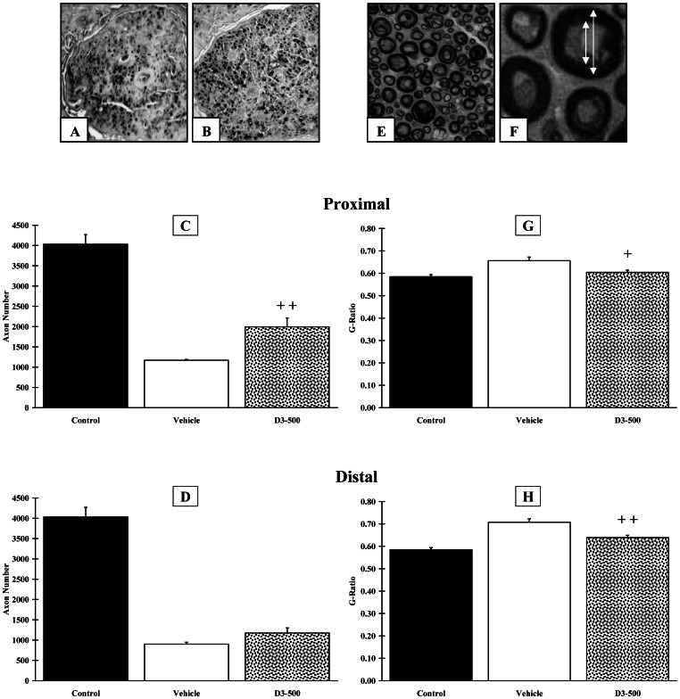
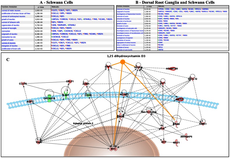
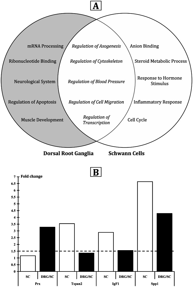
Previously, we demonstrated i) that ergocalciferol (vitamin D2) increases axon diameter and potentiates nerve regeneration in a rat model of transected peripheral nerve and ii) that cholecalciferol (vitamin D3) improves breathing and hyper-reflexia in a rat model of paraplegia. However, before bringing this molecule to the clinic, it was of prime importance i) to assess which form - ergocalciferol versus cholecalciferol - and which dose were the most efficient and ii) to identify the molecular pathways activated by this pleiotropic molecule. The rat left peroneal nerve was cut out on a length of 10 mm and autografted in an inverted position. Animals were treated with either cholecalciferol or ergocalciferol, at the dose of 100 or 500 IU/kg/day, or excipient (Vehicle), and compared to unlesioned rats (Control). Functional recovery of hindlimb was measured weekly, during 12 weeks, using the peroneal functional index. Ventilatory, motor and sensitive responses of the regenerated axons were recorded and histological analysis was performed. In parallel, to identify the genes regulated by vitamin D in dorsal root ganglia and/or Schwann cells, we performed an in vitro transcriptome study. We observed that cholecalciferol is more efficient than ergocalciferol and, when delivered at a high dose (500 IU/kg/day), cholecalciferol induces a significant locomotor and electrophysiological recovery. We also demonstrated that cholecalciferol increases i) the number of preserved or newly formed axons in the proximal end, ii) the mean axon diameter in the distal end, and iii) neurite myelination in both distal and proximal ends. Finally, we found a modified expression of several genes involved in axogenesis and myelination, after 24 hours of vitamin supplementation. Our study is the first to demonstrate that vitamin D acts on myelination via the activation of several myelin-associated genes. It paves the way for future randomised controlled clinical trials for peripheral nerve or spinal cord repair.
Conflict of interest statement
Figures
Similar articles
-
[Repairing the spinal cord with vitamin D: a promising strategy].Biol Aujourdhui. 2014;208(1):69-75. doi: 10.1051/jbio/2014008. Epub 2014 Jun 23. Biol Aujourdhui. 2014. PMID: 24948020 Review. French.
-
Vitamin D2 potentiates axon regeneration.J Neurotrauma. 2008 Oct;25(10):1247-56. doi: 10.1089/neu.2008.0593. J Neurotrauma. 2008. PMID: 18986226
-
Vitamin D3 potentiates myelination and recovery after facial nerve injury.Eur Arch Otorhinolaryngol. 2015 Oct;272(10):2815-23. doi: 10.1007/s00405-014-3305-y. Epub 2014 Sep 27. Eur Arch Otorhinolaryngol. 2015. PMID: 25261104
-
Laminin polymer treatment accelerates repair of the crushed peripheral nerve in adult rats.Acta Biomater. 2019 Mar 1;86:185-193. doi: 10.1016/j.actbio.2019.01.024. Epub 2019 Jan 16. Acta Biomater. 2019. PMID: 30660008 Free PMC article.
-
Peripheral Nerve Regeneration and Muscle Reinnervation.Int J Mol Sci. 2020 Nov 17;21(22):8652. doi: 10.3390/ijms21228652. Int J Mol Sci. 2020. PMID: 33212795 Free PMC article. Review.
Cited by
-
Regenerative Drug Discovery Using Ear Pinna Punch Wound Model in Mice.Pharmaceuticals (Basel). 2022 May 16;15(5):610. doi: 10.3390/ph15050610. Pharmaceuticals (Basel). 2022. PMID: 35631437 Free PMC article.
-
Vitamin D as a potential therapy in amyotrophic lateral sclerosis.CNS Neurosci Ther. 2014 Feb;20(2):101-11. doi: 10.1111/cns.12204. CNS Neurosci Ther. 2014. PMID: 24428861 Free PMC article. Review.
-
Vitamin D as a modulator of pain and inflammation in postmenopausal females with burning mouth syndrome.J Oral Facial Pain Headache. 2025 Mar;39(1):93-102. doi: 10.22514/jofph.2025.008. Epub 2025 Mar 12. J Oral Facial Pain Headache. 2025. PMID: 40129426 Free PMC article.
-
Vitamin D and Its Potential Interplay With Pain Signaling Pathways.Front Immunol. 2020 May 28;11:820. doi: 10.3389/fimmu.2020.00820. eCollection 2020. Front Immunol. 2020. PMID: 32547536 Free PMC article. Review.
-
Experimental immunological demyelination enhances regeneration in autograft-repaired long peripheral nerve gaps.Sci Rep. 2016 Dec 23;6:39828. doi: 10.1038/srep39828. Sci Rep. 2016. PMID: 28008990 Free PMC article.
References
-
- Chabas JF, Alluin O, Rao G, Garcia S, Lavaut MN, et al. (2008) Vitamin D2 potentiates axon regeneration. J Neurotrauma 25: 1247–1256. - PubMed
-
- Fernandes de Abreu DA, Eyles D, Feron F (2009) Vitamin D, a neuro-immunomodulator: implications for neurodegenerative and autoimmune diseases. Psychoneuroendocrinology 34 Suppl 1S265–277. - PubMed
-
- Eyles DW, Smith S, Kinobe R, Hewison M, McGrath JJ (2005) Distribution of the vitamin D receptor and 1 alpha-hydroxylase in human brain. J Chem Neuroanat 29: 21–30. - PubMed
-
- Langub MC, Herman JP, Malluche HH, Koszewski NJ (2001) Evidence of functional vitamin D receptors in rat hippocampus. Neuroscience 104: 49–56. - PubMed
-
- Stumpf WE, O'Brien LP (1987) 1,25 (OH)2 vitamin D3 sites of action in the brain. An autoradiographic study. Histochemistry 87: 393–406. - PubMed
Publication types
MeSH terms
Substances
LinkOut - more resources
Full Text Sources
Other Literature Sources
Medical
