Pyomyositis of the iliacus muscle and pyogenic sacroiliitis after sacroiliac joint block -A case report-
- PMID: 23741573
- PMCID: PMC3668112
- DOI: 10.4097/kjae.2013.64.5.464
Pyomyositis of the iliacus muscle and pyogenic sacroiliitis after sacroiliac joint block -A case report-
Abstract
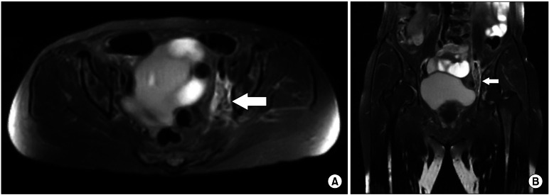
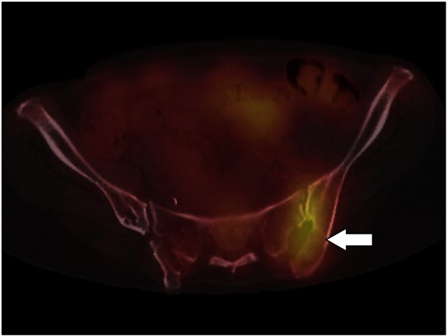
Sacroiliac joint block can be performed for the diagnosis and treatment of sacroiliac joint dysfunction. Although sacroiliac joint block is a common procedure, complications have not been reported in detail. We report a case of iliacus pyomyositis and sacroiliac joint infection following a sacroiliac joint block. A 70-year-old female patient received sacroiliac joint blocks to relieve pelvic pain. The patient was admitted to the emergency room two days after the final sacroiliac joint block (SIJB) with the chief complaints of left pelvic pain corresponding to a visual analogue scale (VAS) score of 9 and fever. A pelvic MRI indicated a diagnosis of myositis. After 1 month of continuous antibiotic therapy, the patient's erythrocyte sedimentation rate (ESR) and C-reactive protein (CRP) level remained elevated. A (67)Ga SPECT/CT was done. Abnormal uptake was seen at the left sacroiliac joint (SIJ), and septic sacroiliitis was suspected. The CRP normalized to 0.29 mg/dl and the ESR decreased to 60 mm/hr, and the patient had no fever after 57 days of antibiotic therapy. She was directed for follow up at an outpatient clinic.
Keywords: Iliacus; Infection; Pyomyositis; Sacroiliac.
Figures
References
-
- Dalin G, Jeffcott LB. Sacroiliac joint of the horse. 1. Gross morphology. Anat Histol Embryol. 1986;15:80–94. - PubMed
-
- Pool-Goudzwaard A, Hoek van, Mulder P, Spoor C, Snijders C, Stoeckart R. The iliolumbar ligament: its influence on stability of the sacroiliac joint. Clin Biomech (Bristol, Avon) 2003;18:99–105. - PubMed
-
- Slipman CW, Whyte WS, 2nd, Chow DW, Chou L, Lenrow D, Ellen M. Sacroiliac joint syndrome. Pain Physician. 2001;4:143–152. - PubMed
-
- Bernard TN, Jr, Kirkaldy-Willis WH. Recognizing specific characteristics of nonspecific low back pain. Clin Orthop Relat Res. 1987;217:266–280. - PubMed
LinkOut - more resources
Full Text Sources
Other Literature Sources
Research Materials
Miscellaneous