

Biomimetic modification of metallic cardiovascular biomaterials: from function mimicking to endothelialization in vivo
- PMID: 23741611
- PMCID: PMC3363017
- DOI: 10.1098/rsfs.2011.0126
Biomimetic modification of metallic cardiovascular biomaterials: from function mimicking to endothelialization in vivo
Abstract
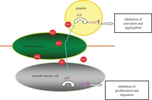
Biosystem-surface interactions play an important role in various biological events and determine the ultimate functionality of implanted devices. Endothelialization or mimicking of endothelium on the surface of cardiovascular materials is a promising way to solve the problems of material-induced thrombosis and restenosis. Meanwhile, a multifunctional surface design is needed as antithrombotic properties should be considered in the period when the implants are not yet completely endothelialized. In this article, we summarize some successful approaches used in our laboratory for constructing multifunctional endothelium-like surfaces on metallic cardiovascular biomaterials through chemical modification of the surface or by the introduction of specific biological molecules to induce self-endothelialization in vivo. Some directions on future research in these areas are also presented.
Keywords: biomimetic modification; endothelial cell; endothelialization; nitric oxide.
Figures
Similar articles
-
Endothelialization of implanted cardiovascular biomaterial surfaces: the development from in vitro to in vivo.J Biomed Mater Res A. 2014 Oct;102(10):3754-72. doi: 10.1002/jbm.a.35025. Epub 2013 Nov 16. J Biomed Mater Res A. 2014. PMID: 24243819 Review.
-
Endothelialization of cardiovascular devices.Acta Biomater. 2019 Nov;99:53-71. doi: 10.1016/j.actbio.2019.08.042. Epub 2019 Aug 24. Acta Biomater. 2019. PMID: 31454565 Review.
-
Surface modification of implanted cardiovascular metal stents: from antithrombosis and antirestenosis to endothelialization.J Biomed Mater Res A. 2014 Feb;102(2):588-609. doi: 10.1002/jbm.a.34714. Epub 2013 Aug 24. J Biomed Mater Res A. 2014. PMID: 23520056 Review.
-
Biomimetic modification of poly(vinyl alcohol): Encouraging endothelialization and preventing thrombosis with antiplatelet monotherapy.Acta Biomater. 2019 Mar 1;86:291-299. doi: 10.1016/j.actbio.2019.01.008. Epub 2019 Jan 10. Acta Biomater. 2019. PMID: 30639349 Free PMC article.
-
Direct surface modification of metallic biomaterials via tyrosine oxidation aiming to accelerate the re-endothelialization of vascular stents.J Biomed Mater Res A. 2018 Feb;106(2):491-499. doi: 10.1002/jbm.a.36258. Epub 2017 Oct 24. J Biomed Mater Res A. 2018. PMID: 28975703
Cited by
-
Natural Biopolymers as Smart Coating Materials of Mesoporous Silica Nanoparticles for Drug Delivery.Pharmaceutics. 2023 Jan 29;15(2):447. doi: 10.3390/pharmaceutics15020447. Pharmaceutics. 2023. PMID: 36839771 Free PMC article. Review.
-
[Construction of controllable polyethylene glycol bioactive coating with hemocompatibility from the surface of modified glass substrate].Sheng Wu Yi Xue Gong Cheng Xue Za Zhi. 2019 Apr 25;36(2):260-266. doi: 10.7507/1001-5515.201705032. Sheng Wu Yi Xue Gong Cheng Xue Za Zhi. 2019. PMID: 31016943 Free PMC article. Chinese.
-
Stabilization and Sterilization of Pericardial Scaffolds by Ultraviolet and Low-Energy Electron Irradiation.Tissue Eng Part C Methods. 2018 Dec;24(12):717-729. doi: 10.1089/ten.TEC.2018.0285. Tissue Eng Part C Methods. 2018. PMID: 30412035 Free PMC article.
-
Accelerating in situ endothelialisation of cardiovascular bypass grafts.Int J Mol Sci. 2014 Dec 29;16(1):597-627. doi: 10.3390/ijms16010597. Int J Mol Sci. 2014. PMID: 25551605 Free PMC article. Review.
-
In situ fabrication of cerium-incorporated hydroxyapatite/magnetite nanocomposite coatings with bone regeneration and osteosarcoma potential.Nanoscale Adv. 2023 Aug 10;5(18):5054-5076. doi: 10.1039/d3na00235g. eCollection 2023 Sep 12. Nanoscale Adv. 2023. PMID: 37705779 Free PMC article.
References
-
- Barnes K. 2008. Complications in patients with ventricular assist devices. Dimens. Crit. Care Nurs. 27, 233–24110.1097/01.DCC.0000338867.24293.20 (doi:10.1097/01.DCC.0000338867.24293.20) - DOI - DOI - PubMed
-
- Ruel M., Kulik A., Lam B. K., Rubens F. D., Hendry P. J., Masters R. G., Bédard P., Mesana T. G. 2005. Long-term outcomes of valve replacement with modern prostheses in young adults. Eur. J. Cardiothorac. Surg. Mar. 27, 425–43310.1016/j.ejcts.2004.12.002 (doi:10.1016/j.ejcts.2004.12.002) - DOI - DOI - PubMed
-
- Cheung Y. F., Sanatani S., Leung M. P., Human D. G., Chau A. K. T., Culham J. A. G. 2000. Early and intermediate-term complications of self-expanding stents limit its potential application in children with congenital heart disease. J. Am. Coll. Cardiol. 35, 1007–101510.1016/S0735-1097(99)00644-0 (doi:10.1016/S0735-1097(99)00644-0) - DOI - DOI - PubMed
-
- Clark D. J., Lessio S., O'Donoghue M., Tsalamandris C., Schainfeld R., Rosenfield K. 2006. Mechanisms and predictors of carotid artery stent restenosis: a serial intravascular ultrasound study. J. Am. Coll. Cardiol. 47, 2390–239610.1016/j.jacc.2006.01.076 (doi:10.1016/j.jacc.2006.01.076) - DOI - DOI - PubMed
-
- Chang C. C., Ong E. T. 2005. Coronary restenosis. Acta. Cardiol. Sin. 21, 177–189
LinkOut - more resources
Full Text Sources
