Review
. 2013 Jun 6:10:182.
doi: 10.1186/1743-422X-10-182.

The modulation of apoptosis by oncogenic viruses

Affiliations
Review

The modulation of apoptosis by oncogenic viruses

Alma Mariana Fuentes-González et al. Virol J. .

Abstract

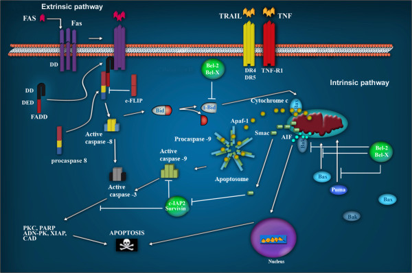
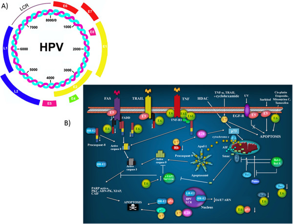
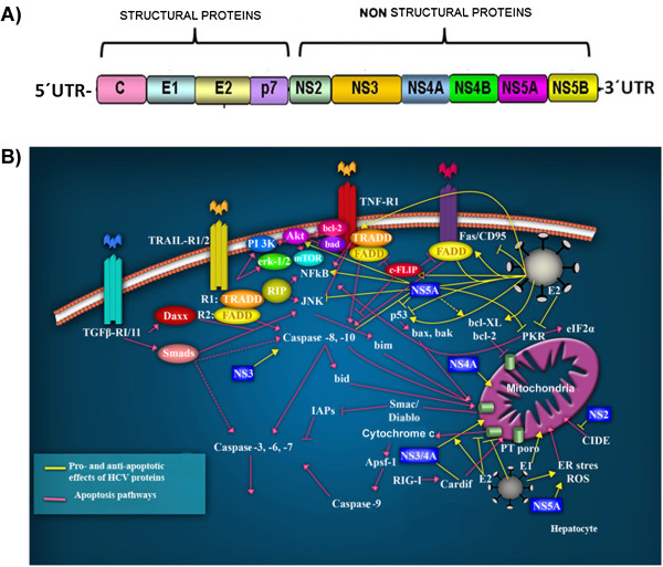
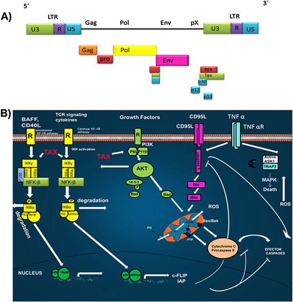
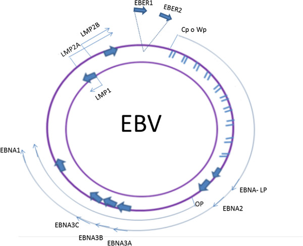
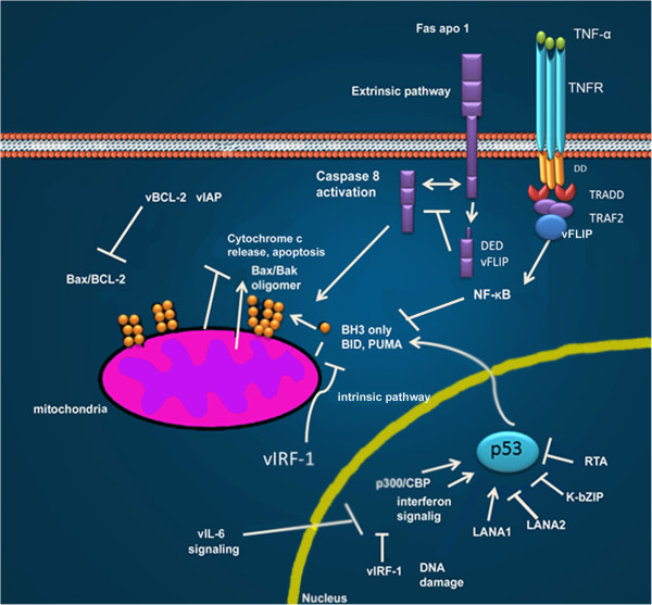
Transforming viruses can change a normal cell into a cancer cell during their normal life cycle. Persistent infections with these viruses have been recognized to cause some types of cancer. These viruses have been implicated in the modulation of various biological processes, such as proliferation, differentiation and apoptosis. The study of infections caused by oncogenic viruses had helped in our understanding of several mechanisms that regulate cell growth, as well as the molecular alterations leading to cancer. Therefore, transforming viruses provide models of study that have enabled the advances in cancer research. Viruses with transforming abilities, include different members of the Human Papillomavirus (HPV) family, Hepatitis C virus (HCV), Human T-cell Leukemia virus (HTLV-1), Epstein Barr virus (EBV) and Kaposi's Sarcoma Herpesvirus (KSHV).Apoptosis, or programmed cell death, is a tightly regulated process that plays an important role in development and homeostasis. Additionally, it functions as an antiviral defense mechanism. The deregulation of apoptosis has been implicated in the etiology of diverse diseases, including cancer. Oncogenic viruses employ different mechanisms to inhibit the apoptotic process, allowing the propagation of infected and damaged cells. During this process, some viral proteins are able to evade the immune system, while others can directly interact with the caspases involved in apoptotic signaling. In some instances, viral proteins can also promote apoptosis, which may be necessary for an accurate regulation of the initial stages of infection.


Figures

Figure 1
Figure 1
Apoptotic signaling pathways. The extrinsic pathway is regulated by membrane receptors. The interaction with their ligands Fas, Trail and TNF, favors their trimerization, inducing the recruitment of FADD through the interaction with their death domains (DDs). The interaction of FADD with procaspase 8 forms a complex called DISC, which favors its oligomerization and auto-cleavage. Active caspase 8 initiates the cascade of effector caspases 3, 6 and 7. In the intrinsic pathway, Bax and Puma are translocated from the cytosol to the mitochondrial membrane as a result of DNA damage, thus provoking the release of cytochrome c. Cytochrome c participates in the formation of the apoptosome, which is involved in DNA degradation. AIF contribute to DNA and nuclear fragmentation.
Figure 2
Figure 2
The HPV proteins involved in apoptotic signaling pathways. A) The Human Papillomavirus Virus genome. All HPVs have a common genomic organization and encode 8 proteins: E1, E2, E4, E5, E6 and E7 (early) and L1 and L2 (late). B) Participation of HPV proteins in apoptotic pathways. E5 impairs the formation of the death-inducing signaling complex triggered by FasL and TRAIL. E6 targets pro-apoptotic proteins such as p53, Bax and Bak for proteolytic degradation; in contrast, E6 can also induce the expression of IAPs. E7 promotes the degradation of the anti-apoptotic protein, pRb, releasing E2F-1. E2 induces apoptosis via the downregulation of E6/E7 mRNA; or direct binding and activation of procaspase 8; or when it binds to p53. Modified from Lagunas-Martínez A. et al. (2010) [49].
Figure 3
Figure 3
The Hepatitis C virus and apoptotic signaling pathways. A) The Hepatitis C virus genome. A single open reading frame encodes four structural proteins and six nonstructural proteins. B) HCV-infected hepatocytes are recognized by the immune cells, that promote apoptosis via the death receptor ligands, TRAIL, TNFα, CD95 ligand, and TGF-β, as well as granzyme B/perforin (Pink lines). Ligand-induced apoptosis activates caspase-8, whereas activation of caspase-9 occurs via the mitochondrial permeability transition (PT) pore, triggering the activation of caspases cascade and the irreversible induction of apoptosis. For virtually all HCV proteins, pro- and anti-apoptotic effects have been described (Yellow lines). The structural (core C, E1 and E2) and nonstructural (NS2, NS3, NS4A and NS5A) proteins participate in the extrinsic and intrinsic apoptotic pathways. Modified from Fischer R. et al. (2007) [92].
Figure 4
Figure 4
HTLV-1 and apoptotic signaling pathways. A) The HTLV-1 viral genome. The genome consists of a single- positive strand of RNA. The long terminal repeats (LTRs) flank the ORF (boxes) of the structural (orange, red, yellow and pink) and the nonstructural (blue, green) viral proteins. B) Main death pathways controlled by HTLV-1 proteins. In infected cells receptor-mediated death (through CD95/Fas) is inhibited by a Tax/NF-kB-mediated upregulation of c-FLIP and IAPs. The PI3K-AKT pathway, also activated by Tax, acts by inhibiting the pro-apoptotic protein Bad and by activating the NF-kB pathway. The accessory proteins, p12 and p13, regulate Bcl-2 and caspases 3 and 9, and also ROS production by mitochondria.
Figure 5
Figure 5
EBV genome. Location and transcription of the EBV latent genes on the double-stranded viral DNA episome. The bottom arrows represent coding exons for each of the latent proteins; the latent proteins include the six nuclear antigens (EBNAs 1, 2, 3A, 3B and 3C, and EBNA-LP) and the three latent membrane proteins (LMPs 1, 2A, 2B). EBNA-LP is transcribed from variable numbers of repetitive exons. The arrows with the discontinuous lines close together at the top represent the highly transcribed nonpolyadenylated RNAs EBER1 and EBER2; their transcription is a consistent feature of latent EBV infection. EBNAs are transcribed from either the Cp or Wp promoter.
Figure 6
Figure 6
Different KSHV proteins inhibit intrinsic and extrinsic apoptotic pathways. vFLIP directly binds to death effector domains, or to the TRAF complex, inhibiting activated-Fas signaling, or activating NF-kB. Both vIAP and vBCL-2 act at the mitochondria to stabilize the mitochondrial membrane and inhibit the activating effects of BH3-only pro-apoptotic molecules.vCyclin LANA1, LANA2, K-bZIP and RTA inhibit p53-induced apoptosis either through direct binding or through inhibition of the p300/CBP coactivator used in p53 transcriptional signaling. vIRFbinds and inhibits pro-death activities of proteins Bid and Bim. Modified from Moore P.S. et al. (2007) [164].

Similar articles

Cited by

References

    1. McLaughlin-Drubin M, Munger K. Viruses associated with human cancer. Biochim Biophys Acta. 2008;1782(suppl 3):127–150. - PMC - PubMed
    1. The Nobel prizes. http://www.nobelprize.org.
    1. zur Hausen H. Viruses in human cancers. Science. 1991;254:1167–1173. - PubMed
    1. Rous P. A Transmissible Avian Neoplasm. (Sarcoma of the Common Fowl.) J Exp Med. 1910;12:696–705. - PMC - PubMed
    1. Shope RE, Hurst EW. Infectious Papillomatosis of Rabbits: With a Note on the Histopathology. J Exp Med. 1933;58:607–624. - PMC - PubMed

Publication types

MeSH terms

LinkOut - more resources