Review
. 2013 Nov;280(22):5570-81.
doi: 10.1111/febs.12373. Epub 2013 Jun 28.

Structure and mechanism of non-histone protein acetyltransferase enzymes

Affiliations
Review

Structure and mechanism of non-histone protein acetyltransferase enzymes

David R Friedmann et al. FEBS J. 2013 Nov.

Abstract

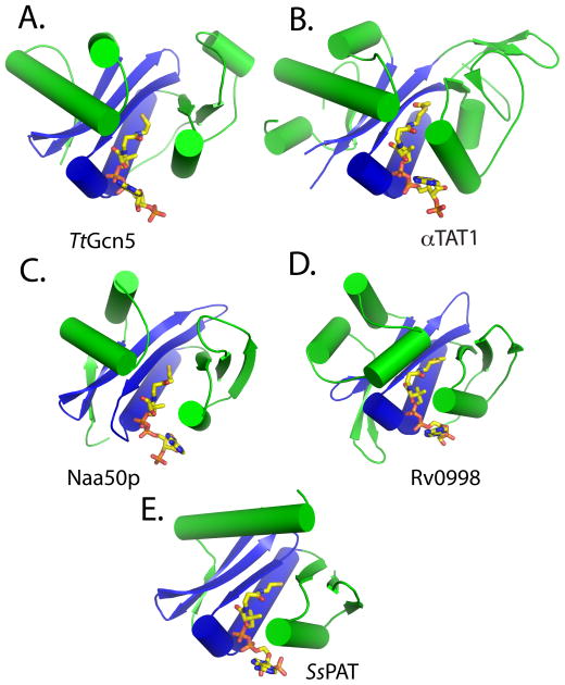
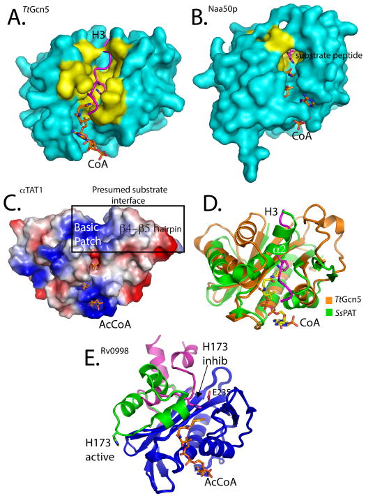
Post-translational modification of proteins is ubiquitous and mediates many cellular processes, including intracellular localization, protein-protein interactions, enzyme activity, transcriptional regulation and protein stability. While the role of phosphorylation as a key post-translational modification has been well studied, the more evolutionarily conserved post-translational modification acetylation has only recently attracted attention as a key regulator of cellular events. Protein acetylation has been largely studied in the context of its role in histone modification and gene regulation, where histones are modified by histone acetyltransferases to promote transcription. However, more recent acetylomic and biochemical studies have revealed that acetylation is mediated by a broader family of protein acetyltransferases. The recent structure determination of several protein acetyltransferases has provided a wealth of molecular information regarding structural features of protein acetyltransferases, their enzymatic mechanisms, their mode of substrate-specific recognition and their regulatory elements. In this review, we briefly describe what is known about non-histone protein substrates, but mainly focus on a few recent structures of protein acetyltransferases to compare and contrast them with histone acetyltransferases to better understand the molecular basis for protein recognition and modification by this family of protein modification enzymes.

Keywords: AcCoA; HATs; NATs; PATs; enzyme mechanism; epigenetics; post-translational modification enzymes; protein acetyltransferases; structure; substrate-binding.


Figures

Figure 1
Figure 1. Structure of non-histone protein acetyltransferases
Cartoon representation of (A) TtGcn5 HAT, (B) αTAT1, (C), Naa50p, (D) activated Rv0998 (only acetyltransferase domain shown), and (E) SsPAT. The conserved acetyltransferase core region is colored blue, and the variable flanking N- and C-terminal segments are colored green. Ac-CoA and CoA are in stick representation and colored according to element: carbon, yellow; nitrogen, blue; oxygen, red.
Fig 2
Fig 2. Catalytic pockets of non-histone protein acetyltransferases
Cartoon representation of acetyltransferases highlighting key catalytic residues colored in cyan and other important residues in yellow. Active site pockets are shown for (A) TtGcn5 with protein contact residues highlighted in yellow, (B) SsPAT, (C) Naa50p with protein contact residues highlighted in yellow, (D) Rv0998, with mutationally sensitive residues in the active site highlighted in yellow and (E) αTAT1 with mutationally sensitive residues in the active site highlighted in yellow. The coloring scheme of the catalytic core and flanking domains is the same as Figure 1.
Figure 3
Figure 3. Substrate specificity determinants and regulation of non-histone protein acetyltransferases
(A) Surface representation of the TtGcn5/CoA/H3 complex. The H3 peptide is represented as a magenta ribbon. Residues described in the text and shown in Figure 2 to interact with the substrate peptide are colored yellow. (B) Surface representation of the Naa50p/CoA/substrate peptide complex also highlighting the peptide substrate binding residues in yellow. (C) Electrostatic surface potential mapping of the αTAT1/AcCoA complex, with surfaces colored according to charge potential (blue-positive, red-negative, white-neutral). The mutationally sensitive and presumed substrate interface is highlighted, emphasizing the large basic patch and location of the β4-β5 hairpin. (D) Carbon alpha alignment of TtGcn5 (orange) and SsPAT (green). The superimposed H3 peptide substrate from the TtGcn5 structure shown in magenta. The α2 helix of SsPAT that overlaps with the H3 peptide-binding site is highlighted. (E) Cartoon representation carbon alpha alignment of the Rv0998 acetyltransferase in the inhibited (magenta) and activated (green) forms. Structurally similar regions are colored blue. The large domain conformational change that results in the movement of residue H173 is highlighted.

References

    1. Walsh C. Posttranslational Modification Of Proteins: Expanding Nature’s Inventory. Roberts and Company Publishers; 2006.
    1. Cohen P. The origins of protein phosphorylation. Nature Cell Biol. 2002;4:E127–30. - PubMed
    1. Cohen P. Protein kinases--the major drug targets of the twenty-first century? Nature Rev Drug Dis. 2002;1:309–15. - PubMed
    1. Kouzarides T. Acetylation: a regulatory modification to rival phosphorylation? EMBO J. 2000;19:1176–9. - PMC - PubMed
    1. Yuan H, Marmorstein R. Histone acetyltransferases: Rising ancient counterparts to protein kinases. Biopolymers. 2013;99:98–111. - PMC - PubMed