Review
. 2013 Aug;280(16):3734-54.
doi: 10.1111/febs.12375. Epub 2013 Jul 5.

Engineering RNA-binding proteins for biology

Affiliations
Review

Engineering RNA-binding proteins for biology

Yu Chen et al. FEBS J. 2013 Aug.

Abstract

RNA-binding proteins play essential roles in the regulation of gene expression. Many have modular structures and combine relatively few common domains in various arrangements to recognize RNA sequences and/or structures. Recent progress in engineering the specificity of the PUF class RNA-binding proteins has shown that RNA-binding domains may be combined with various effector or functional domains to regulate the metabolism of targeted RNAs. Designer RNA-binding proteins with tailored sequence specificity will provide valuable tools for biochemical research as well as potential therapeutic applications. In this review, we discuss the suitability of various RNA-binding domains for engineering RNA-binding specificity, based on the structural basis for their recognition. We also compare various protein engineering and design methods applied to RNA-binding proteins, and discuss future applications of these proteins.

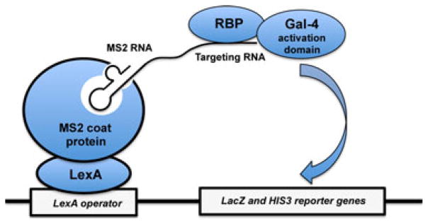
Keywords: RNA recognition motif; RNA-binding domains; RNA-binding proteins; computational design; in vitro evolution; phage display; protein engineering; protein-RNA interactions; yeast three-hybrid system; zinc finger.


Figures

Fig. 1
Fig. 1
Engineered RNA-binding proteins may be combined with various functional domains for diverse biological applications. The RNA-binding domains (RBDs) are shown as horizontal arrays of green rectangles containing multiple linked domains, but the number of RBDs varies according to the protein and the required RNA-binding affinity and specificity. (A) Targeted translation regulation. Engineered RBDs may be linked to a translational activator (e.g. GLD2) or a translational repressor (e.g. CAF1) to affect the translation and stability of specific mRNAs in vivo [23]. (B) Regulation of alternative splicing. Sequence-specific RNA-binding domains may be combined with various splicing modulatory domains to specifically regulate different types of alternative splicing events [43]; for example, an arginine/serine-rich (RS) domain from ASF/SF2 may be used as a splicing activator and a glycine-rich (Gly) domain from hnRNP A1 may be used as a splicing repressor. (C) Imaging RNA in living cells. Engineered RBDs may be tethered with fluorescent proteins (e.g. GFP) for real-time imaging of RNA trafficking in living cells [20]. (D) Control of RNA localization. Engineered RBDs may be fused to a nuclear localization signal (NLS) to direct the RNA to the nucleus [16]. (E) Cleavage or degradation of specific RNAs. Sequence-specific RBDs may be fused to a non-specific RNase to cut or degrade a target RNA [22]. (F) Targeted inhibitors of RNA functions. Engineered RBDs may be simply used to target a specific sequence of RNA to sterically block its function through RNA conformational changes or inhibition of its interactions with other functional proteins [14].
Fig 2
Fig 2
Structures of RNA-recognition motifs (RRMs) in complex with ssRNAs. The proteins are shown as ribbons, colored according to their secondary structures (red, helices; yellow, sheets; green, loops). Conserved aromatic residues on protein binding surfaces are highlighted. All the structural figures in the paper were prepared using PyMOL [122]. The RNAs are shown as blue or cyan stick models. (A, B) Single RRM recognition: crystal structure of the U1A spliceosomal protein complexed with an RNA hairpin (PDB entry 1URN) (A) and solution structure of the RBD of Fox–1 in complex with UGCAUGU (PDB entry 2ERR) (B). (C, D) Multiple RRMs: crystal structure of an AU-rich element recognized by the HuD protein (PDB entry 1G2E) (C) and crystal structure of the poly(A)-binding protein in complex with polyadenylate RNA (PDB entry 1CVJ) (D).
Fig. 3
Fig. 3
RNA recognition by PUF proteins (adapted from Chen et al. 2011). The crystal structure shows an engineered PUF domain in complex with RNA (PDB entry 2YJY). The image on the right is a close-up view of the RNA recognition code of PUF repeats for each nucleotide in the structure.
Fig. 4
Fig. 4
Structures of KH domains in complex with RNA. (A) Crystal structure of the Nova KH domain bound to RNA (PDB entry 1EC6). (B) Two KH domains from the NusA protein form a large interaction surface to bind RNA in an extended conformation (PDB entry 2ASB).
Fig. 5
Fig. 5
RNA binding by ZF proteins. (A) Crystal structure of a three-finger polypeptide from TIFIIIA (4–6) in complex with truncated 5S RNA (61 nt) (PDB entry 1UN6). (b) Solution structure of the RNA complex of TIS11d (PDB entry 1RGO). (C) NMR structure of human Lin28 zinc knuckles bound to the sequence 5′-AGGAGAU-3′ from the pre-let–7 terminal loop region. The structure shows that each zinc knuckle recognizes an AG dinucleotide separated by a single nucleotide spacer (PDB entry 2LI8). (D) Crystal structure of a ZF from ZRANB2 bound to a single-stranded GGU-containing RNA (PDB entry 3G9Y). Protein side chains forming base-specific contacts with GGU are shown in green; zinc ions are shown as magenta spheres.
Fig. 6
Fig. 6
Double-stranded RNA recognition by dsRBDs. (A) Structure of dsRBD1 of ADAR2 bound to USL RNA (PDB entry 2L3C). Helix α1 and the β1–β2 loop that mediate sequence-specific contacts are labeled. (B) Crystal structure of an RNaseIII–RNA complex containing an RNaseIII dimer and two dsRNA molecules (PDB entry 2EZ6). The two dsRBDs are shown as ribbon models, two endonuclease domains (endoND) are shown as a surface model, and the RNA is shown as a stick model.
Fig. 7
Fig. 7
Schematic representation of the yeast three-hybrid system used to screen protein–RNA interactions (adapted from Dong et al. 2011). The interaction between a Gal4-RBP (e.g. Gal4-PUF) and target RNA fused to the MS2 binding sequence triggers expression of both the LacZ and HIS3 reporter genes.
Fig. 8
Fig. 8
Illustration of the strategy used for selection of RBPs by in vitro compartmentalization (adapted from Chen et al. 2008). RBPs are fused to an N–terminal ZF DNA-binding polypeptide that recognizes a cognate sequence present in multiple copies upstream of the coding region in the linear DNA templates. The library of ZF-RBP genes in aqueous phase is emulsified and compartmentalized in water-in-oil droplets, and the corresponding fusion protein is expressed by a coupled in vitro transcription/translation reaction. The expressed chimeric proteins bind to their encoding DNA templates through the zinc fingers. After breaking the emulsions, streptavidin-coated magnetic beads bound with biotin-labeled RNA are used to capture the RBPs and corresponding encoding DNA; the selected gene expression cassettes are subsequently amplified by PCR.

References

    1. Chen Y, Varani G. Protein families and RNA recognition. FEBS J. 2005;272:2088–2097. - PubMed
    1. Lunde BM, Moore C, Varani G. RNA-binding proteins: modular design for efficient function. Nat Rev Mol Cell Biol. 2007;8:479–490. - PMC - PubMed
    1. Pavletich NP, Pabo CO. Zinc finger–DNA recognition: crystal structure of a Zif268–DNA complex at 2.1 Å. Science. 1991;252:809–817. - PubMed
    1. Klug A. The discovery of zinc fingers and their applications in gene regulation and genome manipulation. Annu Rev Biochem. 2010;79:213–231. - PubMed
    1. Jamieson AC, Miller JC, Pabo CO. Drug discovery with engineered zinc-finger proteins. Nat Rev Drug Discov. 2003;2:361–368. - PubMed

MeSH terms

Substances