Review
. 2013 Jun 6:8:90.
doi: 10.1186/1746-1596-8-90.

Solitary myofibroma of the sigmoid colon: case report and review of the literature

Affiliations
Review

Solitary myofibroma of the sigmoid colon: case report and review of the literature

Mi-Jung Kim et al. Diagn Pathol. .

Abstract

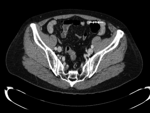
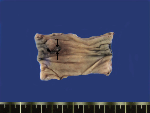
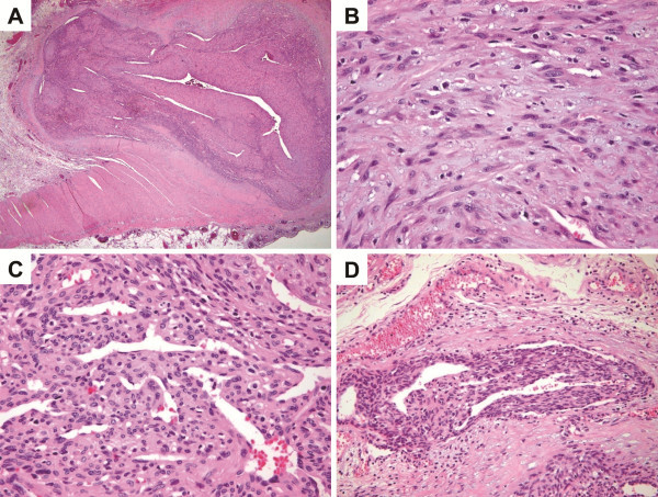
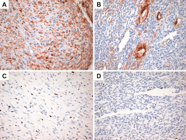
A 58-year-old woman presented with a solitary myofibroma that arose in the sigmoid colon. Computed tomography revealed a highly enhanced intramural mass (1.3-cm maximum diameter) in the proximal sigmoid colon. Histologically, the tumor exhibited a biphasic growth pattern, which comprised haphazardly arranged, interwoven fascicles of plump, myoid-appearing spindle cells with elongated nuclei and abundant eosinophilic cytoplasm, and more cellular areas of primitive-appearing polygonal cells that were arranged in a hemangiopericytomatous pattern. The tumor cells were positive for smooth muscle actin (SMA), and negative for desmin, h-caldesmon, CD34, cytokeratin, S100 protein, and CD117. The Ki-67 labeling index was not high (up to 7%). Based on these histologic and immunohistochemical features, our patient was diagnosed with a myofibroma of the sigmoid colon. The presence of solitary myofibroma in the intestine of an adult requires attention to avoid misdiagnosis as a more aggressive mesenchymal tumor.

Virtual slides: The virtual silde(s) for this article can be found here: http://www.diagnosticpathology.diagnomx.eu/vs/2096403796957687.


Figures

Figure 1
Figure 1
Radiologic characterization of the lesion. CT imaging revealed a highly enhanced intramural mass (1.3 cm in maximum diameter; white arrows) in the proximal sigmoid colon.
Figure 2
Figure 2
Gross features of the lesion. A well-demarcated, ovoid 1.3 cm × 1.0 cm × 0.7 cm intramural mass (arrows) is noted in the colon.
Figure 3
Figure 3
Histologic features of the tumor. A. The tumor is well-demarcated and situated transmurally. There are alternating light and dark areas within the tumor (H&E, ×20). B. The light area consists of haphazardly arranged, interweaving fascicles of plump, myoid-appearing spindle cells with elongated nuclei and abundant eosinophilic cytoplasm (H&E, ×400). C. The dark, more cellular area consists of primitive-appearing polygonal cells arranged in a hemangiopericytomatous pattern (H&E, ×400). D. Intravascular proliferation of tumor cells is noted at the periphery of the lesion (H&E, ×200).
Figure 4
Figure 4
Immunohistochemical findings. A. The myoid-appearing spindle tumor cells show diffuse and strong positivity for SMA (immunostaining, ×400). B. The primitive-appearing tumor cells are focally positive for SMA (immunostaining, ×400). C. The myoid-appearing spindle tumor cells show an increased Ki-67 labeling index (up to 7%) (immunostaining, ×400). D. The primitive-appearing tumor cells display low Ki-67 labeling index (up to 1%) (immunostaining, ×400).

References

    1. Chung EB, Enzinger FM. Infantile myofibromatosis. Cancer. 1981;48:1807–1818. doi: 10.1002/1097-0142(19811015)48:8<1807::AID-CNCR2820480818>3.0.CO;2-G. - DOI - PubMed
    1. Fletcher CDM, Unni KK, Mertens F. World health organization classifiction of tumors. Pathology and genetics of tumors of soft tissue and bone. Lyon: IARC Press; 2002.
    1. Marks MK, Dewan PA, Stokes KB, Smith AL, McKelvie P. Infantile myofibromatosis causing biliary and pancreatic obstruction: a case report. Med Pediatr Oncol. 1988;16:363–365. doi: 10.1002/mpo.2950160515. - DOI - PubMed
    1. Schurr P, Moulsdale W. Infantile myofibroma: a case report and review of the literature. Adv Neonatal Care. 2008;8:13–20. doi: 10.1097/01.ANC.0000311012.63887.fb. Review. - DOI - PubMed
    1. Fine SW, Davis NJ, Lykins LE, Montgomery E. Solitary testicular myofibroma: a case report and review of the literature. Arch Pathol Lab Med. 2005;129:1322–1325. Review. - PubMed

Substances

LinkOut - more resources