

Relative stability of different DNA guanine quadruplex stem topologies derived using large-scale quantum-chemical computations
- PMID: 23742743
- PMCID: PMC3775466
- DOI: 10.1021/ja402525c
Relative stability of different DNA guanine quadruplex stem topologies derived using large-scale quantum-chemical computations
Abstract
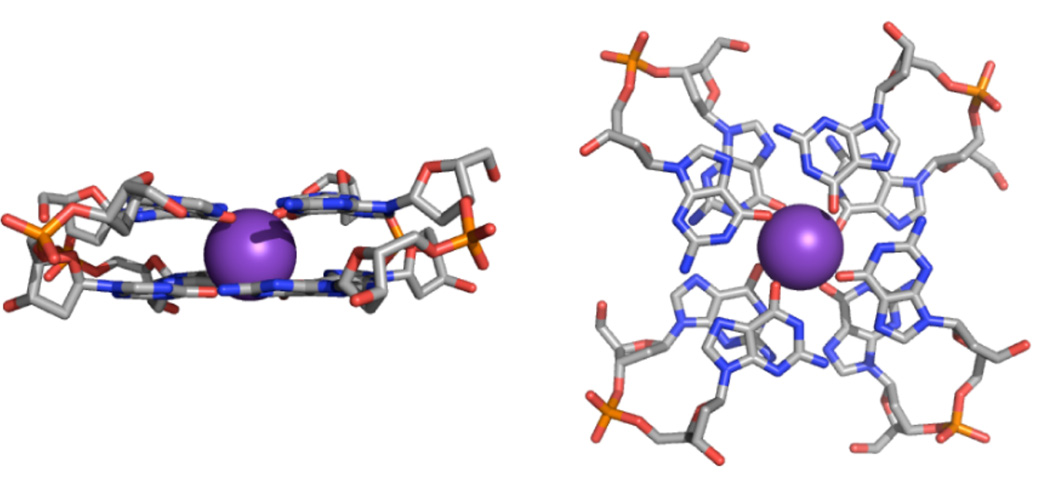
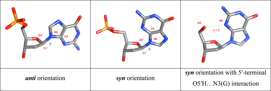
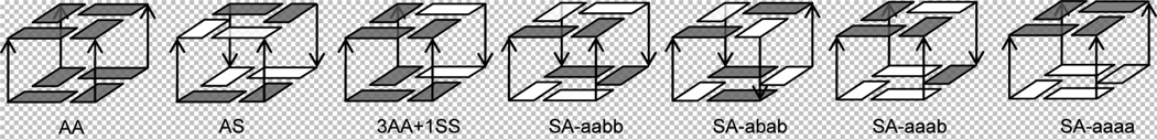
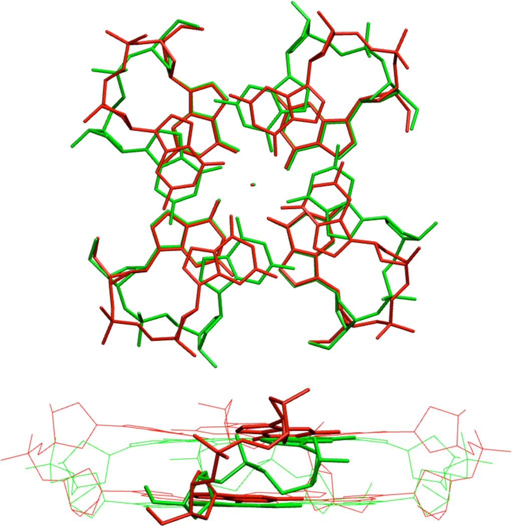
We provide theoretical predictions of the intrinsic stability of different arrangements of guanine quadruplex (G-DNA) stems. Most computational studies of nucleic acids have applied Molecular Mechanics (MM) approaches using simple pairwise-additive force fields. The principle limitation of such calculations is the highly approximate nature of the force fields. In this study, we for the first time apply accurate QM computations (DFT-D3 with large atomic orbital basis sets) to essentially complete DNA building blocks, seven different folds of the cation-stabilized two-quartet G-DNA stem, each having more than 250 atoms. The solvent effects are approximated by COSMO continuum solvent. We reveal sizable differences between MM and QM descriptions of relative energies of different G-DNA stems, which apparently reflect approximations of the DNA force field. Using the QM energy data, we propose correction to earlier free energy estimates of relative stabilities of different parallel, hybrid, and antiparallel G-stem folds based on classical simulations. The new energy ranking visibly improves the agreement between theory and experiment. We predict the 5'-anti-anti-3' GpG dinucleotide step to be the most stable one, closely followed by the 5'-syn-anti-3' step. The results are in good agreement with known experimental structures of 2-, 3-, and 4-quartet G-DNA stems. Besides providing specific results for G-DNA, our study highlights basic limitations of force field modeling of nucleic acids. Although QM computations have their own limitations, mainly the lack of conformational sampling and the approximate description of the solvent, they can substantially improve the quality of calculations currently relying exclusively on force fields.
Figures
Similar articles
-
Derivation of Reliable Geometries in QM Calculations of DNA Structures: Explicit Solvent QM/MM and Restrained Implicit Solvent QM Optimizations of G-Quadruplexes.J Chem Theory Comput. 2016 Apr 12;12(4):2000-16. doi: 10.1021/acs.jctc.5b01025. Epub 2016 Mar 8. J Chem Theory Comput. 2016. PMID: 26914292
-
Can We Execute Reliable MM-PBSA Free Energy Computations of Relative Stabilities of Different Guanine Quadruplex Folds?J Phys Chem B. 2016 Mar 24;120(11):2899-912. doi: 10.1021/acs.jpcb.6b01059. Epub 2016 Mar 11. J Phys Chem B. 2016. PMID: 26918369
-
Benchmark quantum-chemical calculations on a complete set of rotameric families of the DNA sugar-phosphate backbone and their comparison with modern density functional theory.Phys Chem Chem Phys. 2013 May 21;15(19):7295-310. doi: 10.1039/c3cp44383c. Phys Chem Chem Phys. 2013. PMID: 23575975
-
The MOD-QM/MM Method: Applications to Studies of Photosystem II and DNA G-Quadruplexes.Methods Enzymol. 2016;577:443-81. doi: 10.1016/bs.mie.2016.05.021. Epub 2016 Jul 15. Methods Enzymol. 2016. PMID: 27498648 Free PMC article. Review.
-
How to understand quantum chemical computations on DNA and RNA systems? A practical guide for non-specialists.Methods. 2013 Nov;64(1):3-11. doi: 10.1016/j.ymeth.2013.05.025. Epub 2013 Jun 7. Methods. 2013. PMID: 23747334 Review.
Cited by
-
Small Heterocyclic Ligands as Anticancer Agents: QSAR with a Model G-Quadruplex.Molecules. 2022 Nov 4;27(21):7577. doi: 10.3390/molecules27217577. Molecules. 2022. PMID: 36364401 Free PMC article.
-
Balancing the interactions of ions, water, and DNA in the Drude polarizable force field.J Phys Chem B. 2014 Jun 19;118(24):6742-57. doi: 10.1021/jp503469s. Epub 2014 Jun 9. J Phys Chem B. 2014. PMID: 24874104 Free PMC article.
-
Comprehensive analysis of intramolecular G-quadruplex structures: furthering the understanding of their formalism.Nucleic Acids Res. 2024 Apr 24;52(7):3522-3546. doi: 10.1093/nar/gkae182. Nucleic Acids Res. 2024. PMID: 38512075 Free PMC article.
-
Quantum Mechanical Investigation of the G-Quadruplex Systems of Human Telomere.ACS Omega. 2018 Aug 27;3(8):9934-9944. doi: 10.1021/acsomega.8b01678. eCollection 2018 Aug 31. ACS Omega. 2018. PMID: 31459122 Free PMC article.
-
B-DNA Structure and Stability: The Role of Nucleotide Composition and Order.ChemistryOpen. 2022 Feb;11(2):e202100231. doi: 10.1002/open.202100231. Epub 2022 Jan 27. ChemistryOpen. 2022. PMID: 35083880 Free PMC article.
References
-
- Cornell WD, Cieplak P, Bayly CI, Gould IR, Merz KM, Ferguson DM, Spellmeyer DC, Fox T, Caldwell JW, Kollman PA. J. Am. Chem. Soc. 1995;117:5179–5197.
-
- Foloppe N, MacKerell AD. J. Comput. Chem. 2000;21:86–104.
-
- Mackerell AD. J. Comput. Chem. 2004;25:1584–1604. - PubMed
Publication types
MeSH terms
Substances
Grants and funding
LinkOut - more resources
Full Text Sources
Other Literature Sources
Miscellaneous
