

Diagnostic ability of retinal nerve fiber layer maps to detect localized retinal nerve fiber layer defects
- PMID: 23743523
- PMCID: PMC3772356
- DOI: 10.1038/eye.2013.119
Diagnostic ability of retinal nerve fiber layer maps to detect localized retinal nerve fiber layer defects
Abstract
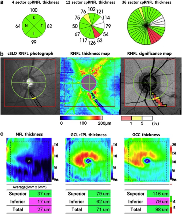
Purpose: To evaluate and compare the diagnostic ability of spectral domain optical coherence tomography (SD-OCT) for detecting localized retinal nerve fiber layer (RNFL) defects in topographic RNFL maps and circumpapillary RNFL (cpRNFL) thickness measurements.
Methods: Sixty-four eyes with localized RNFL defects in red-free RNFL photographs and 72 healthy eyes were included. All participants were imaged with SD-OCT. The area and angular width of the localized RNFL defects were measured with ImageJ software on RNFL thickness map, significance map (yellow pixels, <5% level), and red-free RNFL photographs. The sensitivity, specificity, and area under the receiver operating characteristic curves (AUCs) were calculated for cpRNFL thickness, macular inner retina thickness, and RNFL maps (thickness, significance) according to the quantitative measurements and a <5% level of classification to distinguish eyes with localized RNFL defects from healthy eyes.
Results: RNFL thickness map (sensitivity 96.9-98.4%, specificity 86.1-98.6%, and AUCs 0.915-0.992) and significance map (sensitivity 96.9-98.4%, specificity 88.9-95.8%, and AUCs 0.937-0.983) showed superior performance in detecting localized RNFL defects compared with other parameters (P-value 0.001-0.024) except for 36 sector cpRNFL thickness (sensitivity 92.2%, specificity 87.5%, and AUCs 0.898; P-value 0.080-0.545). The sensitivity for detecting RNFL defects was related to the angular width, area, and depth of the RNFL defects in the cpRNFL (4 sector, 12 sector) and macular inner retinal measurements. RNFL thickness and significance maps showed a constant sensitivity regardless of variations in angular width, area, and depth of the RNFL defects.
Conclusion: RNFL thickness and significance maps could be used to distinguish eyes with localized RNFL defects from healthy eyes more effectively than cpRNFL thickness and macular inner retina thickness measurements.
Figures
References
-
- Huang ML, Chen HY. Development and comparison of automated classifiers for glaucoma diagnosis using Stratus optical coherence tomography. Invest Ophthalmol Vis Sci. 2005;46:4121–4129. - PubMed
-
- Parikh RS, Parikh S, Sekhar GC, Kumar RS, Prabakaran S, Babu JG, et al. Diagnostic capability of optical coherence tomography (Stratus OCT 3) in early glaucoma. Ophthalmology. 2007;114:2238–2243. - PubMed
-
- Ye C, To E, Weinreb RN, Yu M, Liu S, Lam DS, et al. Comparison of retinal nerve fiber layer imaging by spectral domain optical coherence tomography and scanning laser ophthalmoscopy. Ophthalmology. 2011;118:2196–2202. - PubMed
-
- Leung CK, Lam S, Weinreb RN, Liu S, Ye C, Liu L, et al. Retinal nerve fiber layer imaging with spectral-domain optical coherence tomography: analysis of the retinal nerve fiber layer map for glaucoma detection. Ophthalmology. 2010;117:1684–1691. - PubMed
Publication types
MeSH terms
LinkOut - more resources
Full Text Sources
Other Literature Sources
Medical
