. 2013 Jul 16;110(29):12000-5.
doi: 10.1073/pnas.1301278110. Epub 2013 Jun 6.

An MLL-dependent network sustains hematopoiesis

Affiliations

An MLL-dependent network sustains hematopoiesis

Erika L Artinger et al. Proc Natl Acad Sci U S A. .

Abstract

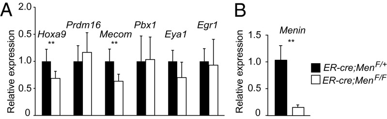
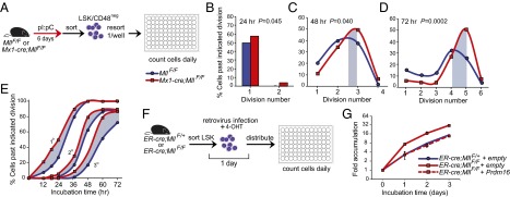
The histone methyltransferase Mixed Lineage Leukemia (MLL) is essential to maintain hematopoietic stem cells and is a leukemia protooncogene. Although clustered homeobox genes are well-characterized targets of MLL and MLL fusion oncoproteins, the range of Mll-regulated genes in normal hematopoietic cells remains unknown. Here, we identify and characterize part of the Mll-dependent transcriptional network in hematopoietic stem cells with an integrated approach by using conditional loss-of-function models, genomewide expression analyses, chromatin immunoprecipitation, and functional rescue assays. The Mll-dependent transcriptional network extends well beyond the previously appreciated Hox targets, is comprised of many characterized regulators of self-renewal, and contains target genes that are both dependent and independent of the MLL cofactor, Menin. Interestingly, PR-domain containing 16 emerged as a target gene that is uniquely effective at partially rescuing Mll-deficient hematopoietic stem and progenitor cells. This work highlights the tissue-specific nature of regulatory networks under the control of MLL/Trithorax family members and provides insight into the distinctions between the participation of MLL in normal hematopoiesis and in leukemia.

Keywords: HSC; epigenetics; proliferation.


Conflict of interest statement

Conflict of interest statement: P.E. is a shareholder of Amgen stock.

Figures

Fig. 1.
Fig. 1.
Identification of Mll-regulated genes in HSCs. General overview of genes up-regulated (A) or down-regulated (B) in Mll-deficient LSK/CD48neg cells compared with controls. Cells were sorted from pI:pC-injected control MllF/F or Mx1-cre;MllF/F animals at day six. Gene Ontology assignments were based on the criteria in Datasets S1 and S2. (C) The top down-regulated transcription factors in Mll-deficient LSK/CD48neg cells listed by fold reduction (see also Dataset S2). (D) RT-qPCR validating down-regulated genes in independent control MllF/F (blue) or Mll-deficient (red) LSK/CD48neg cells, n = 8 animals per genotype; ND, not detected. (E) RT-qPCR validation of transcripts in LSK cells sorted from control ER-cre;MllF/+ (blue) or ER-cre;MllF/F animals (red) cultured for 72 h after initiating Mll deletion. Relative expression levels were determined by normalizing to Gapdh, n = 4 animals per genotype. Error bars represent 95% confidence interval (CI). *P ≤ 0.07, **P ≤ 0.05. ER-cre, estrogen receptorT2 mutant fused to cre recombinase.
Fig. 2.
Fig. 2.
MLL binds directly to the promoter regions of a subset of genes identified by expression array. ChIP results demonstrating specific enrichment at the Mecom locus (Mds1 and Evi1 start sites) and the Prdm16, Pbx1, and Eya1 promoter regions. Anti-MLL C-terminal (black) or control (anti-GAL4, gray) antibodies were used for ChIP, and enrichment was determined by using quantitative PCR assays. Amplicon position is indicated relative to the TSS for each gene. Results using additional primers surrounding the TSS are shown in Fig. S4. Data represents averages ± SEM for two to four PCR replicates and are representative of at least four independent experiments.
Fig. 3.
Fig. 3.
Menin loss affects some but not all MLL targets in LSK cells. (A) RT-qPCR of Mll-regulated genes in LSK cells sorted from control ER-cre;Men1F/+ (black) or ER-cre;Men1F/F cells (white) cultured for 72 h after initiating Menin deletion. Expression levels were normalized to rRNA. (B) Menin transcript levels in LSK cells treated as in A. Error bars represent 95% CI; n = 4–8 animals per genotype. **P ≤ 0.05.
Fig. 4.
Fig. 4.
Effect of reexpression of individual Mll targets on others in the network. RT-qPCR of genes in LSK cells reexpressing the cDNA indicated below each set of bars. Cells were produced in vivo by pI:pC injection, sorted 6 d later, then infected with a retrovirus without an added cDNA (empty) or cDNA as indicated. Two days later, retrovirally infected cells were sorted and RT-qPCR assays were performed. (A) Prdm16 expression levels in control MllF/F (blue) or Mll-deficient (red) LSK cells infected with the retrovirus indicated below each set of bars. Expression levels were normalized to the average expression level empty retrovirus-infected MllF/F cells and to Gapdh in each sample. Expression of Hoxa9 (B), Mecom transcripts (C), Pbx1 (D), and Eya1 (E) were analyzed and normalized as in A. Dashed lines indicate the average expression level in wild-type or Mll-deficient, empty retrovirus infected cells; four to five animals per genotype were used for each experiment, and error bars represent 95% CI. P values are shown for the comparison between pairs of empty vector and Evi1-expressing cells, calculated with the paired Student t test. ND, not detected.
Fig. 5.
Fig. 5.
Reexpression of Prdm16 partially rescues Mll deficiency. (A) Experimental scheme to determine effects of reexpression of Mll-dependent genes. LSK cells were sorted from control MllF/F or Mx1-cre;MllF/F donor animals then infected with the indicated retrovirus. The entire pool of infected and uninfected cells was transplanted into irradiated recipients, which were analyzed 6 wk later. (B) Results of reexpression of each individual gene in control MllF/F (blue) or Mll-deficient LSK cells (red); each point represents an individual recipient animal, n = 3–10 recipients per condition. The percentage of donor-type (CD45.1+) BM cells that are GFP+ or hCD4+ 2 wk after Mll deletion is shown. Data are representative of three independent experiments. *P ≤ 0.05 was calculated by using the Wilcoxon rank-sum test.
Fig. 6.
Fig. 6.
The intrinsic proliferation defect of Mll-deficient HSCs is corrected by reexpression of Prdm16. (A) Scheme to determine proliferation kinetics of individual LSK/CD48neg cells. Mll deletion was performed in vivo, and double-sorted LSK/CD48neg cells were deposited at 1 cell per well. Cell divisions were scored every 24 h. (BD) Cumulative proliferation data from individual control MllF/F (blue) or Mll-deficient LSK/CD48neg cells (red). Data represent 158 control MllF/F and 240 Mll-deficient cells; n = 3–5 animals per genotype. The difference between modes of each line is indicated by gray fill. The Pearson’s χ2 test was performed to determine statistical significance, shown on BD. (E) Higher-resolution proliferation kinetics of control MllF/F (blue) or Mll-deficient LSK/CD48neg cells (red). Cells were prepared as in A, n = 2–3 animals per genotype, 93 control MllF/F and 38 Mll-deficient cells. The percentage of cells past the first, second, and third divisions are graphed separately (1°, 2°, 3°). (F) Scheme to determine the impact of Prdm16 reexpression in Mll-deficient LSK cells. (G) Accumulation of LSK cells expressing an empty (solid) or hCD4-Prdm16 retrovirus (dashed). LSK cells were sorted from control ER-cre;MllF/+ (blue) or ER-cre;MllF/F animals (red), cultured in 4-OHT during the retroviral infection to induce Mll deletion then enumerated every 24 h for 3 d. Data represent averages ± 95% CI, n = 4 animals per genotype, 3 replicates per time point.

Comment in

References

    1. Schuettengruber B, Chourrout D, Vervoort M, Leblanc B, Cavalli G. Genome regulation by polycomb and trithorax proteins. Cell. 2007;128(4):735–745. - PubMed
    1. Sparmann A, van Lohuizen M. Polycomb silencers control cell fate, development and cancer. Nat Rev Cancer. 2006;6(11):846–856. - PubMed
    1. Deshpande AJ, Bradner J, Armstrong SA. Chromatin modifications as therapeutic targets in MLL-rearranged leukemia. Trends Immunol. 2012;33(11):563–570. - PMC - PubMed
    1. Daser A, Rabbitts TH. Extending the repertoire of the mixed-lineage leukemia gene MLL in leukemogenesis. Genes Dev. 2004;18(9):965–974. - PubMed
    1. Krivtsov AV, Armstrong SA. MLL translocations, histone modifications and leukaemia stem-cell development. Nat Rev Cancer. 2007;7(11):823–833. - PubMed

Publication types

MeSH terms

Substances

Associated data