

Anti-HIV host factor SAMHD1 regulates viral sensitivity to nucleoside reverse transcriptase inhibitors via modulation of cellular deoxyribonucleoside triphosphate (dNTP) levels
- PMID: 23744077
- PMCID: PMC3711331
- DOI: 10.1074/jbc.M113.472159
Anti-HIV host factor SAMHD1 regulates viral sensitivity to nucleoside reverse transcriptase inhibitors via modulation of cellular deoxyribonucleoside triphosphate (dNTP) levels
Abstract
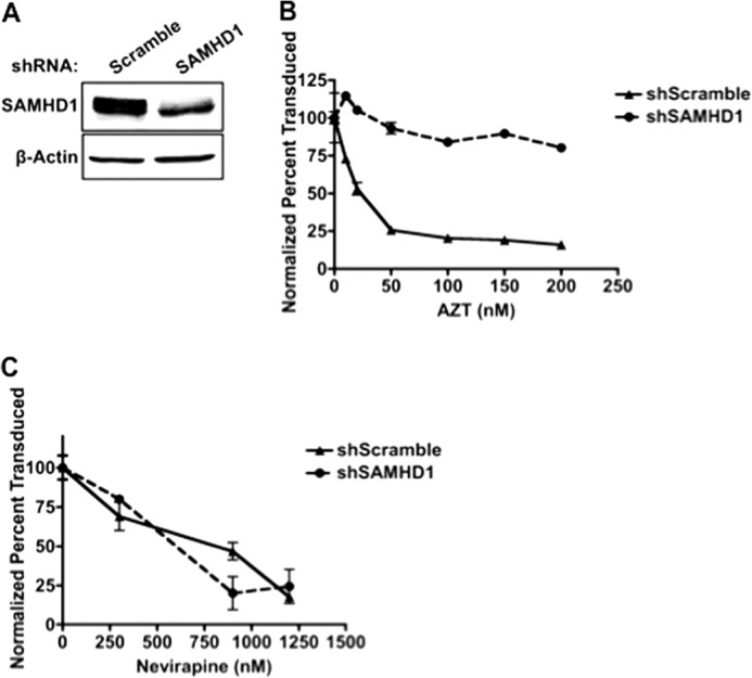
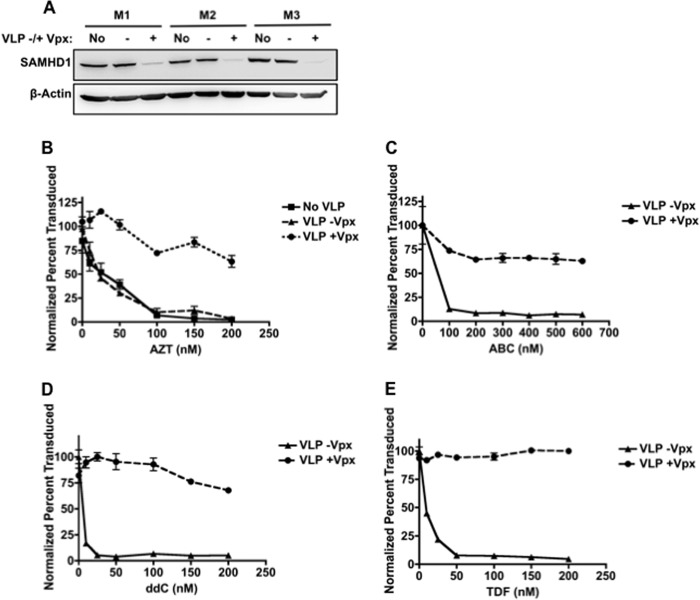
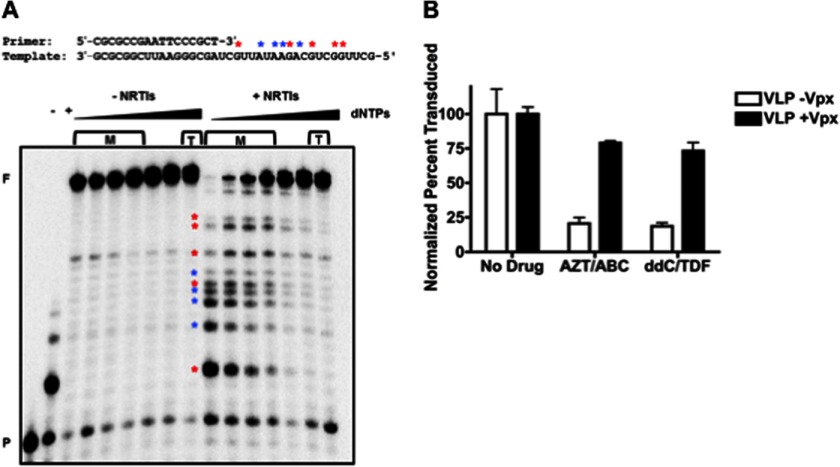
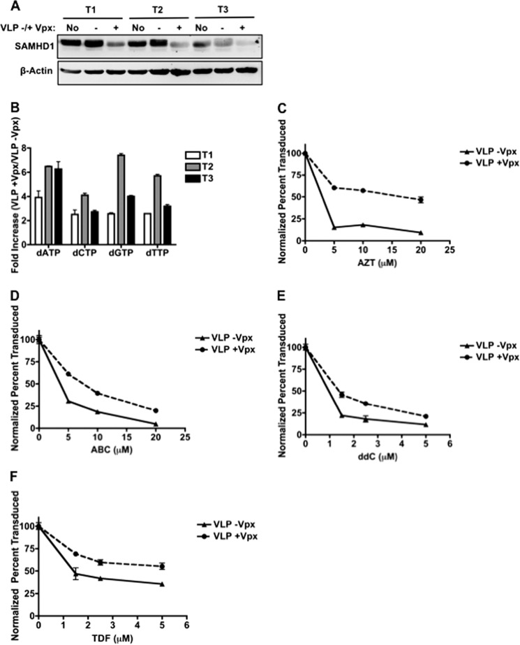
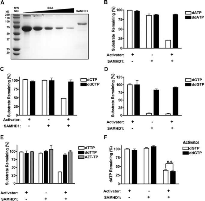
Newly identified anti-HIV host factor, SAMHD1, restricts replication of lentiviruses such as HIV-1, HIV-2, and simian immunodeficiency virus in macrophages by enzymatically hydrolyzing and depleting cellular dNTPs, which are the substrates of viral DNA polymerases. HIV-2 and some simian immunodeficiency viruses express viral protein X (VPX), which counteracts SAMHD1 and elevates cellular dNTPs, enhancing viral replication in macrophages. Because nucleoside reverse transcriptase inhibitors (NRTIs), the most commonly used anti-HIV drugs, compete against cellular dNTPs for incorporation into proviral DNA, we tested whether SAMHD1 directly affects the efficacy of NRTIs in inhibiting HIV-1. We found that reduction of SAMHD1 levels with the use of virus-like particles expressing Vpx- and SAMHD1-specific shRNA subsequently elevates cellular dNTPs and significantly decreases HIV-1 sensitivity to various NRTIs in macrophages. However, virus-like particles +Vpx treatment of activated CD4(+) T cells only minimally reduced NRTI efficacy. Furthermore, with the use of HPLC, we could not detect SAMHD1-mediated hydrolysis of NRTI-triphosphates, verifying that the reduced sensitivity of HIV-1 to NRTIs upon SAMHD1 degradation is most likely caused by the elevation in cellular dNTPs.
Keywords: DNA Polymerase; DNA Replication; HIV-1; Macrophages; Nucleoside Nucleotide Analogs.
Figures
References
-
- Ho D. D., Neumann A. U., Perelson A. S., Chen W., Leonard J. M., Markowitz M. (1995) Rapid turnover of plasma virions and CD4 lymphocytes in HIV-1 infection. Nature 373, 123–126 - PubMed
-
- Clements J. E., Zink M. C., Narayan O., Gabuzda D. H. (1994) Lentivirus infection of macrophages. Immunol. Ser. 60, 589–600 - PubMed
-
- Roshal M., Kim B., Zhu Y., Nghiem P., Planelles V. (2003) Activation of the ATR-mediated DNA damage response by the HIV-1 viral protein R. J. Biol. Chem. 278, 25879–25886 - PubMed
-
- Singh D. K., Chebloune Y., Mselli-Lakhal L., Karr B. M., Narayan O. (1999) Ovine lentivirus-infected macrophages mediate productive infection in cell types that are not susceptible to infection with cell-free virus. J. Gen. Virol. 80, 1437–1444 - PubMed
Publication types
MeSH terms
Substances
Grants and funding
- DE007202/DE/NIDCR NIH HHS/United States
- P30 AI050409/AI/NIAID NIH HHS/United States
- GM049573/GM/NIGMS NIH HHS/United States
- R01 AI077401/AI/NIAID NIH HHS/United States
- R01 GM049573/GM/NIGMS NIH HHS/United States
- GM068411/GM/NIGMS NIH HHS/United States
- T32 GM068411/GM/NIGMS NIH HHS/United States
- T32 DE007202/DE/NIDCR NIH HHS/United States
- T90 DE021985/DE/NIDCR NIH HHS/United States
- R01 AI049781/AI/NIAID NIH HHS/United States
- R01 GM104198/GM/NIGMS NIH HHS/United States
- R56 AI049781/AI/NIAID NIH HHS/United States
- AI049781/AI/NIAID NIH HHS/United States
- 5P30-AI-50409/AI/NIAID NIH HHS/United States
- AI077401/AI/NIAID NIH HHS/United States
LinkOut - more resources
Full Text Sources
Other Literature Sources
Research Materials
Miscellaneous
