. 2013 Jun 6;4(6):e653.
doi: 10.1038/cddis.2013.185.

A novel Osmium-based compound targets the mitochondria and triggers ROS-dependent apoptosis in colon carcinoma

Affiliations

A novel Osmium-based compound targets the mitochondria and triggers ROS-dependent apoptosis in colon carcinoma

A Maillet et al. Cell Death Dis. .

Abstract

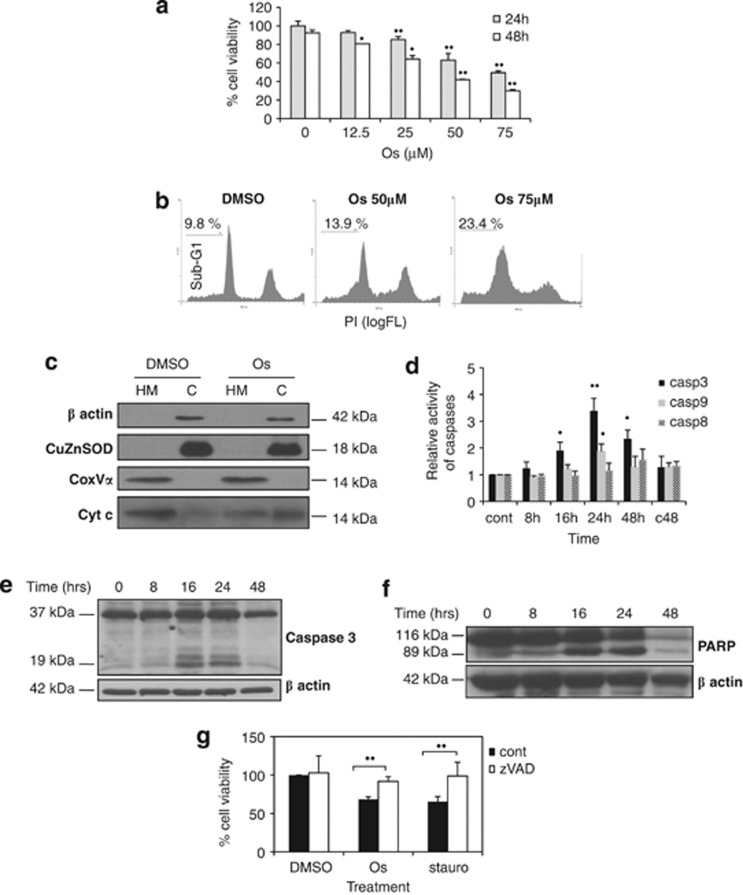
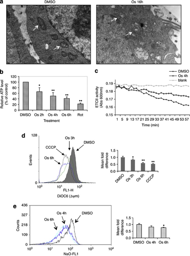
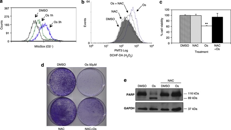
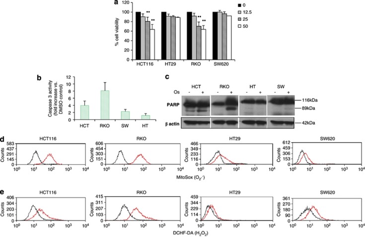
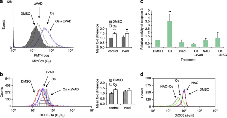
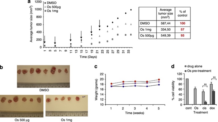
Engagement of the mitochondrial-death amplification pathway is an essential component in chemotherapeutic execution of cancer cells. Therefore, identification of mitochondria-targeting agents has become an attractive avenue for novel drug discovery. Here, we report the anticancer activity of a novel Osmium-based organometallic compound (hereafter named Os) on different colorectal carcinoma cell lines. HCT116 cell line was highly sensitive to Os and displayed characteristic features of autophagy and apoptosis; however, inhibition of autophagy did not rescue cell death unlike the pan-caspase inhibitor z-VAD-fmk. Furthermore, Os significantly altered mitochondrial morphology, disrupted electron transport flux, decreased mitochondrial transmembrane potential and ATP levels, and triggered a significant increase in reactive oxygen species (ROS) production. Interestingly, the sensitivity of cell lines to Os was linked to its ability to induce mitochondrial ROS production (HCT116 and RKO) as HT29 and SW620 cell lines that failed to show an increase in ROS were resistant to the death-inducing activity of Os. Finally, intra-peritoneal injections of Os significantly inhibited tumor formation in a murine model of HCT116 carcinogenesis, and pretreatment with Os significantly enhanced tumor cell sensitivity to cisplatin and doxorubicin. These data highlight the mitochondria-targeting activity of this novel compound with potent anticancer effect in vitro and in vivo, which could have potential implications for strategic therapeutic drug design.


Figures

Figure 1
Figure 1
Os induces cell death in cancer cells via apoptosis. (a) HCT116 cells (1.25 × 10⁵) were seeded in 24-well plates, treated with increasing doses of Os for 24 or 48 h and cell survival was assessed by crystal violet assay. Results are expressed as average of the percentage of the relative growth. HCT116 cells were treated with 50 μM of Os for 16 h, (b) cell cycle profiles were obtained by PI staining and (c) subcellular fractions were subjected to western blot analysis. (d) Os-treated cells were harvested and assayed for caspases 3, 8 and 9 activities using fluorogenic substrates, as described in Materials and Methods. Lysates from cells treated with Os (50 μM) were probed for processing of caspase 3 (e) and PARP cleavage (f). (g) Cells were preincubated with zVAD (100 μM for 1.5 h) before exposure to Os (50 μM) or staurosporine (2.5 μM) for 24 h, and survival was assessed by crystal violet staining. P<0.05, ••P<0.025 versus control
Figure 2
Figure 2
Os affects mitochondrial function and morphology. (a) HCT116 cells were treated with Os (50 μM) for 16 h, fixed and viewed under electron microscope (magnification × 25 000) White arrows pointing to mitochondria. (b) Relative ATP level was determined in HCT116 cells using the ATP Bioluminescence Assay Kit CLS II (Roche), as described in Materials and Methods. (c) A total of 6 × 10⁵ HCT116 cells were incubated with 50 μM of Os for 4 h and ETC-II activity was determined using the Human Complex activity Microplate Assay Kit (Mitosciences). (d) Os-treated cells were incubated with the potential-sensitive probe DiOC6, and ΔΨm was analyzed by flow cytometry as described in Materials and Methods. Representative bar graphs were plotted with the G-mean values to determine the fold difference in ΔΨm between control and Os-treated groups. (e) Os-treated cells were loaded with N-nonyl-acridine-orange, and cardiolipin oxidation was analyzed by flow cytometry as described in Materials and Methods. Quantification of G-mean values have been reported on the corresponding graphs. P<0.05, ••P<0.025 versus control
Figure 3
Figure 3
Os-induced cell death is ROS-dependent. (a) A total of 3 × 10⁵ HCT116 cells were incubated with 50 μM of Os for 1 or 3 h, and intramitochondrial O2 was determined using the fluorescent dye MitoSox Red and analyzed by flow cytometry. (b) Cells were preincubated with 1.5 mM of NAC for 1.5 h before incubation with 50 μM of Os for 4 h, and intracellular H2O2 was detected by DCFH-DA loading and analyzed by flow cytometry. (c) Cells were preincubated with 1.5 mM of NAC for 1.5 h before incubation with 50 μM of Os for 24 h. Cell viability was determined by crystal violet. (d) Cells were treated with NAC and Os as in (b) and 20 000 cells were seeded onto 100-mm petridishes for assessment of colony formation. (e) After preincubation with NAC and Os treatment, cell lysates were used for western blot analysis using PARP antibody. ••P<0.025 versus control
Figure 4
Figure 4
Colon cancer cells producing less ROS are resistant to Os-induced cell death. (a) HCT116, HT29, RKO and SW620 colon cancer cell lines were seeded in 24-well plates, treated with increasing doses of Os for 24 h (12.5–50 μM), and cell survival was assessed by crystal violet assay. (b) Os-treated cells were harvested after 24 h and assayed for caspase 3 activity using fluorogenic substrates. (c) After 24 h of Os treatment (50 μM), cells were harvested and lysates probed for processing of PARP cleavage. (d) HCT116, HT29, RKO and SW620 cells were incubated with 50 μM of Os for 5 h, intramitochondrial O2 was determined using the fluorescent dye MitoSox Red (e) and intracellular H2O2 was detected by DCFH-DA loading (d), and cells were analyzed by flow cytometry. ••P<0.025 versus control
Figure 5
Figure 5
ROS production is upstream of caspases' activation. HCT116 cells were preincubated with zVAD (100 μM for 1.5 h) before exposure to Os (50 μM); intramitochondrial O2 was determined using the fluorescent dye MitoSox Red (a), and intracellular H2O2 was detected by DCFH-DA loading (b) before being analyzed by flow cytometry. Representative bar graphs were plotted with the G-mean values. (c) Cells were pretreated with 100 μM of zVAD or 1.5 mM of NAC for 1.5 h, incubated with Os (50 μM) for 24 h and assayed for caspase 3 activity using fluorogenic substrates. (d) HCT116 cells were preincubated with 1.5 mM of NAC, then treated with 50 μM Os for 5 h and loaded with the potential-sensitive probe DiOC6, and ΔΨm was analyzed by flow cytometry. ••P<0.025 versus control
Figure 6
Figure 6
Os inhibits tumorigenesis in vivo and sensitizes cancer cells to cytotoxic agents. (a) Balb/c nude mice carrying HCT116 ectopic tumors (around 50 mm3) received intraperitoneally Os (500 μg, equivalent to 28 mg/kg), Os (1 mg, equivalent to 56 mg/kg) or DMSO two times a week during 3 weeks. All the mice were killed after 10 more days of monitoring. Arrows indicate the time of injections. The results are presented as average of six animals. (b) After killing, the tumors were collected and measured. NT=no tumor. (c) Mice weight was measured on a weekly basis, results represent the average weight of the six animals per group. Blue: DMSO, purple: Os 1 mg, red: Os 500 μg. (d) HCT116 cells (1.25 × 10⁵) were seeded in 24-well plates and treated with either 50  μM of Os alone, 50 μM of cisplatin alone, 5 μM of doxorubicin alone for 24 h or pretreated with 50  μM of Os for 2 h and then with 50 μM of cisplatin (cis), 5 μM of doxorubicin (dox) for 24 h. Cell survival was assessed by crystal violet assay. ••P<0.025 versus control

References

    1. Tan ML, Ooi JP, Ismail N, Moad AI, Muhammad TS. Programmed cell death pathways and current antitumor targets. Pharm Res. 2009;26:1547–1560. - PubMed
    1. Dias N, Bailly C. Drugs targeting mitochondrial functions to control tumor cell growth. Biochem Pharmacol. 2005;70:1–12. - PubMed
    1. Fulda S, Galluzzi L, Kroemer G. Targeting mitochondria for cancer therapy. Nat Rev Drug Discov. 2010;9:447–464. - PubMed
    1. Modica-Napolitano JS, Kulawiec M, Singh KK. Mitochondria and human cancer. Curr Mol Med. 2007;7:121–131. - PubMed
    1. Neuzil J, Dong LF, Rohlena J, Truksa J, Ralph SJ. Classification of mitocans, anti-cancer drugs acting on mitochondria. Mitochondrion. 2012;13:199–208. - PubMed

Publication types

LinkOut - more resources