. 2013 Jun 6;4(6):e656.
doi: 10.1038/cddis.2013.133.

PARG dysfunction enhances DNA double strand break formation in S-phase after alkylation DNA damage and augments different cell death pathways

Affiliations

PARG dysfunction enhances DNA double strand break formation in S-phase after alkylation DNA damage and augments different cell death pathways

H Shirai et al. Cell Death Dis. .

Abstract

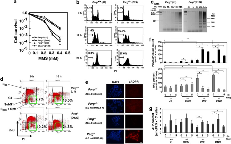
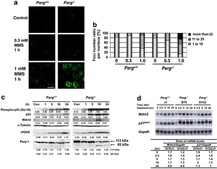
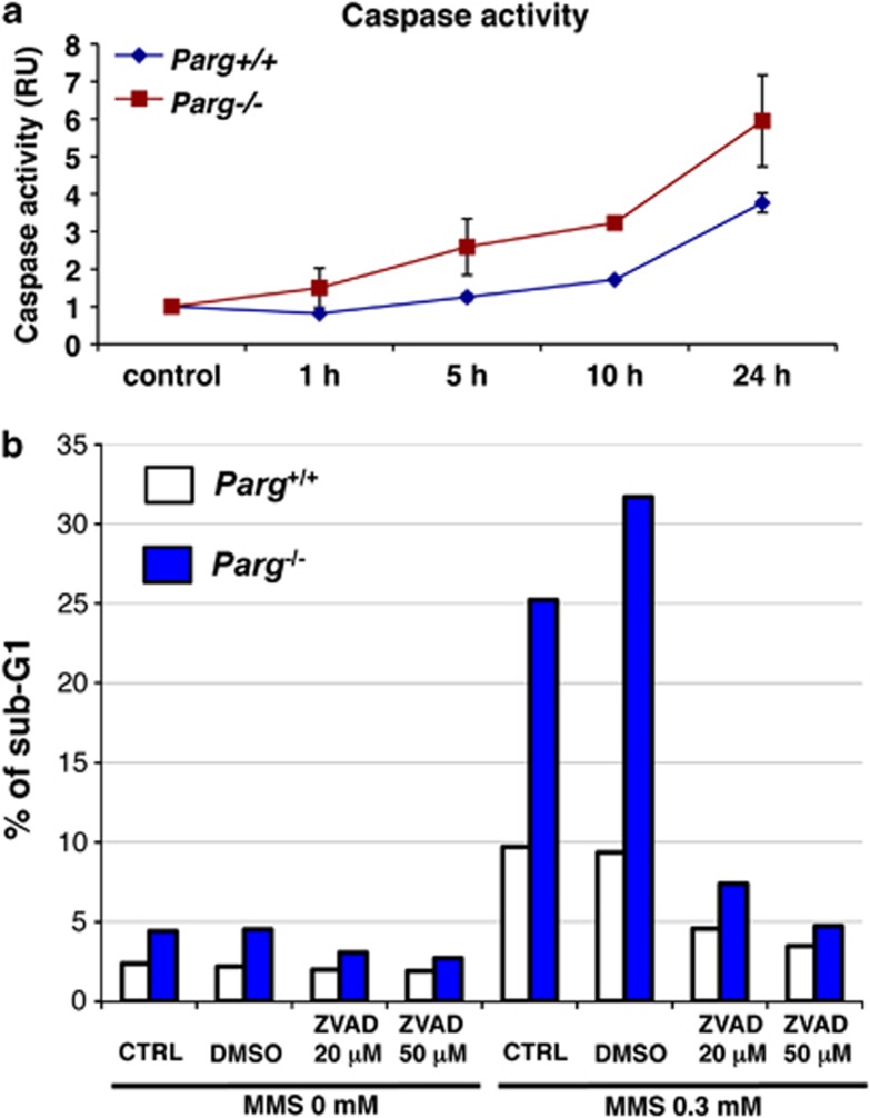
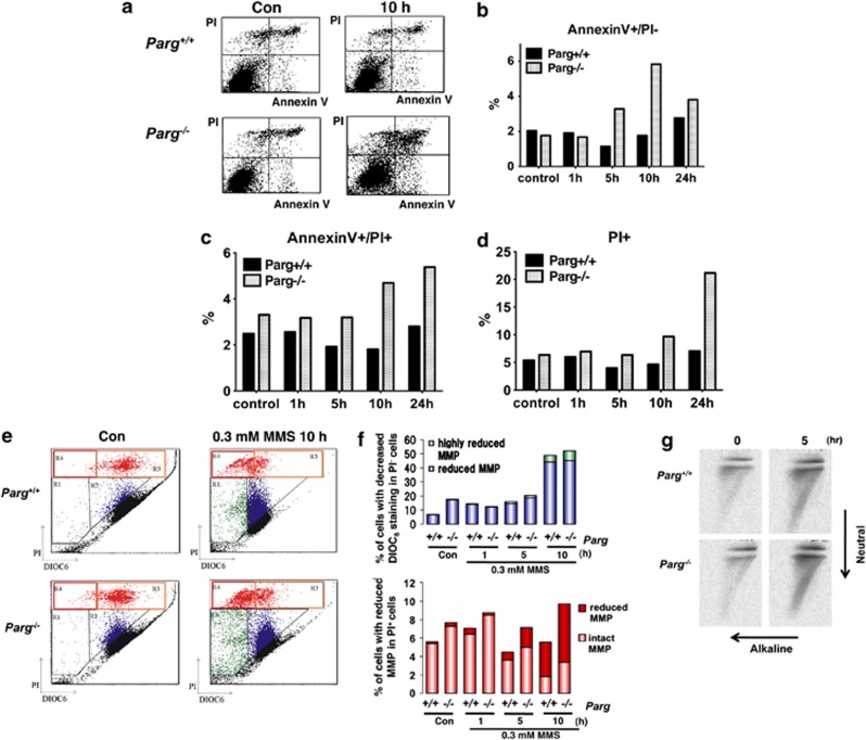
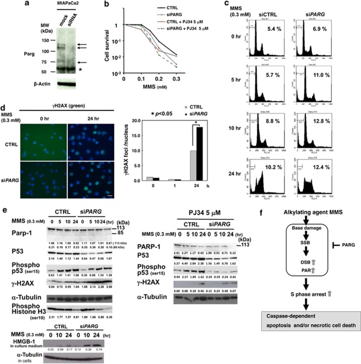
Poly(ADP-ribose) glycohydrolase (PARG) is the primary enzyme responsible for the degradation of poly(ADP-ribose). PARG dysfunction sensitizes cells to alkylating agents and induces cell death; however, the details of this effect have not been fully elucidated. Here, we investigated the mechanism by which PARG deficiency leads to cell death in different cell types using methylmethanesulfonate (MMS), an alkylating agent, and Parg(-/-) mouse ES cells and human cancer cell lines. Parg(-/-) mouse ES cells showed increased levels of γ-H2AX, a marker of DNA double strand breaks (DSBs), accumulation of poly(ADP-ribose), p53 network activation, and S-phase arrest. Early apoptosis was enhanced in Parg(-/-) mouse ES cells. Parg(-/-) ES cells predominantly underwent caspase-dependent apoptosis. PARG was then knocked down in a p53-defective cell line, MIAPaCa2 cells, a human pancreatic cancer cell line. MIAPaCa2 cells were sensitized to MMS by PARG knockdown. Enhanced necrotic cell death was induced in MIAPaCa2 cells after augmenting γ-H2AX levels and S-phase arrest. Taken together, these data suggest that DSB repair defect causing S-phase arrest, but p53 status was not important for sensitization to alkylation DNA damage by PARG dysfunction, whereas the cell death pathway is dependent on the cell type. This study demonstrates that functional inhibition of PARG may be useful for sensitizing at least particular cancer cells to alkylating agents.


Figures

Figure 1
Figure 1
Enhanced cell death in Parg−/− ES cells after MMS treatment. ES cells plated on STO feeder cells were treated with different concentrations of MMS. (a) Clonogenic survival assay. (b) Flow cytometric analysis of the apoptotic sub-G1 population and cell cycle distribution. In the absence of MMS treatment, cell cycle distribution was not altered by Parg deficiency. (c) Time course analysis of DNA ladder formation following the treatment with 0.3 mM MMS. With this method, the fragmented DNA was solely recovered as described in the Materials and Methods. The fragmented DNA obtained from the same number of inoculated cells was subjected to electrophoresis. (d) Two-dimensional flow cytometry analysis of EdU incorporation and PI staining after MMS treatment. SDS corresponds to S-phase fraction showing DNA synthesis. SBDS corresponds to S-phase fraction showing blocked DNA synthesis. Percentage of the cells in SBDS fraction are marked with green line and their percentages are presented. (e) Immunostaining of ES cells with 10H antibody 1 h after treatment with 0.3 mM MMS. Bar 40 μm. (fg) Measurement of PAR and NAD (f), and ATP (g) levels in ES whole-cell extracts after treatment with 0.3 mM MMS. PAR was extracted and fully digested by recombinant GST-Parg, and the amount of ADP-ribose was measured. Asterisks indicate statistically significant differences (P<0.05). Untreated Parg+/+ ES cells contained ∼9 pmol of PAR (measured as the amount of ADP-ribosylated residues) per 5 × 10⁶ cells, which is comparable to that reported in various cell types
Figure 2
Figure 2
Early changes in the DDR after MMS treatment in Parg−/− ES cells. (a) Immunocytochemistry of γH2AX foci formation. Bar 15 μm. (b) The number of foci in ES cells was counted and quantified 1 h after treatment with 0.3 mM MMS. (c) Western blot analysis of p53, mdm2, γH2AX, and Parp-1. Western blot shows enhanced cleavage of Parp-1 in Parg−/− ES cells. The values under the bands show the intensities of the bands normalized by the band of α-tubulin. Whereas caspases cleave Parp-1 to yield an 89-kDa C-terminal fragment, calpain-mediated cleavage of Parp-1 will produce a 40 kDa N-terminal fragment. (d) Northern blot analysis of Mdm2 and p21waf1 expression. The lower panel shows mRNA levels normalized to Gapdh expression
Figure 3
Figure 3
Enhanced caspase activity and suppression of enhanced cell death by caspase inhibition in Parg−/− ES cells, following treatment with 0.3 mM MMS. (a) Caspase activity. (b) Cell death in Parg−/− ES cells in the presence and absence of the caspase inhibitor ZVAD at 20 and 50 μM. Mean values of representative duplicate experiments are plotted
Figure 4
Figure 4
Early and late changes in plasma membrane and mitochondrial membrane potential in ES cells after treatment with 0.3 mM MMS. (a) Time-course analysis of changes in plasma membrane integrity using propidium iodide (PI) and annexin V staining. Percentage of PI-positive (b), Annexin V-positive and PI-negative (c), and annexin V-positive and PI-positive fractions (d). Representative data are shown. (e and f) Mitochondrial membrane potential was measured by DIOC6 staining. (g) Two-dimensional electrophoresis is performed in one dimension under neutral conditions to analyze DSBs, and then in a second dimension under alkaline conditions to analyze single strand lesions. Fragmentation of mitochondrial DNA was then examined using a probe for the mitochondria-specific gene, ND1
Figure 5
Figure 5
Enhanced sensitivity to MMS treatment in MIAPaCa2 induced by PARG knockdown. (a) Western blot analysis of PARG in MIAPaCa2. (b) Clonogenic survival assay of MIAPaCa2 after MMS treatment. We also compared the effect by PARG knockdown with PARP inhibitor PJ-34 at 5 μM and analyzed the combination effects of PARG knockdown and PJ-34. (c) Flow cytometric analysis of cell cycle distribution in MIAPaCa2. Percentages of sub-G1 fraction are shown. (d) Analysis of γH2AX foci formation in MIAPaCa2 after treatment with 0.3 mM MMS. Bar 40 μm. (e) Western blot analysis of proteins involved in the DDR response (left panel) and necrosis (left panel, bottom part) after treatment with 0.3 mM MMS in PARG knockdown MIAPaCa2. Combination effect of PARP inhibitor PJ-34 at 5 μM was also analyzed for DDR response (right panel). The values under the bands show the intensities of the bands normalized by the band of α-tubulin. (f) An induction model of different cell death pathways through common DSB increase and S-phase arrest in p53 active and defective cells. This study implies that p53-dependent response itself is not important for sensitization to alkylation DNA damage by PARG dysfunction

Similar articles

Cited by

References

    1. Sugimura T. Poly(adenosine diphosphate ribose) Prog Nucleic Acid Res Mol Biol. 1973;13:127–151. - PubMed
    1. Miwa M, Masutani M. PolyADP-ribosylation and cancer. Cancer Sci. 2007;98:1528–1535. - PMC - PubMed
    1. Meyer-Ficca ML, Meyer RG, Coyle DL, Jacobson EL, Jacobson MK. Human poly(ADP-ribose) glycohydrolase is expressed in alternative splice variants yielding isoforms that localize to different cell compartments. Exp Cell Res. 2004;297:521–532. - PubMed
    1. Fujihara H, Ogino H, Maeda D, Shirai H, Nozaki T, Kamada N, et al. Poly(ADP-ribose) Glycohydrolase deficiency sensitizes mouse ES cells to DNA damaging agents. Curr Cancer Drug Targets. 2009;9:953–962. - PubMed
    1. Oka S, Kato J, Moss J. Identification and characterization of a mammalian 39-kDa poly(ADP-ribose) glycohydrolase. J Biol Chem. 2006;281:705–713. - PubMed

Publication types

MeSH terms