. 2013 Jun 6;4(6):e662.
doi: 10.1038/cddis.2013.189.

Hyperosmotic stress induces cell cycle arrest in retinal pigmented epithelial cells

Affiliations

Hyperosmotic stress induces cell cycle arrest in retinal pigmented epithelial cells

T Arsenijevic et al. Cell Death Dis. .

Abstract

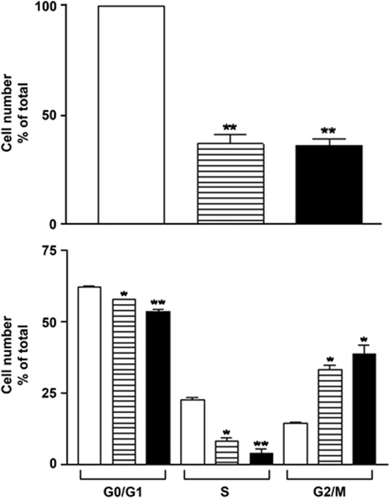
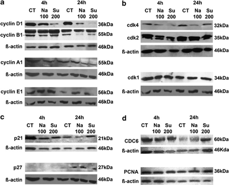
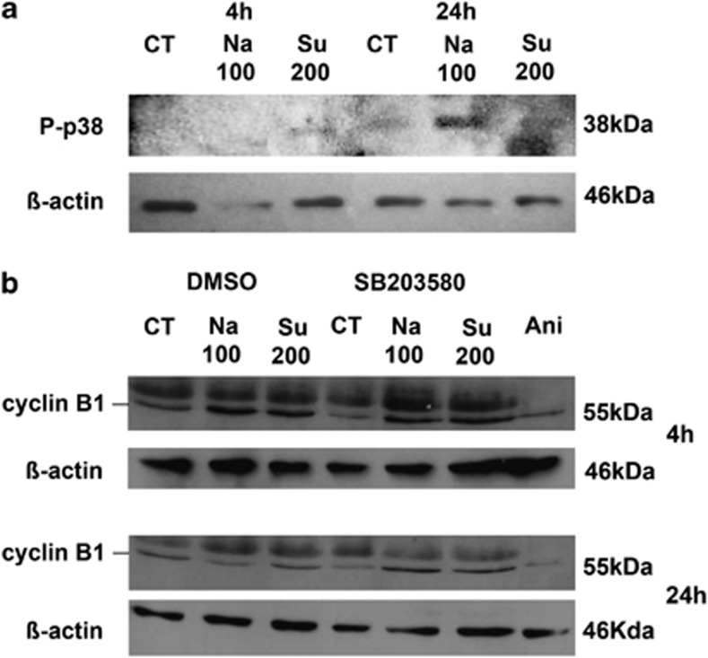
Osmotic changes occur in many tissues and profoundly influence cell function. Herein, we investigated the effect of hyperosmotic stress on retinal pigmented epithelial (RPE) cells using a microarray approach. Upon 4-h exposure to 100 mM NaCl or 200 mM sucrose, 79 genes were downregulated and 72 upregulated. Three gene ontology categories were significantly modulated: cell proliferation, transcription from RNA polymerase II promoter and response to abiotic stimulus. Fluorescent-activated cell sorting analysis further demonstrated that owing to hyperosmotic stimulation for 24 h, cell count and cell proliferation, as well as the percentage of cells in G0/G1 and S phases were significantly decreased, whereas the percentage of cells in G2/M phases increased, and apoptosis and necrosis remained unaffected. Accordingly, hyperosmotic conditions induced a decrease of cyclin B1 and D1 expression, and an activation of the p38 mitogen-activated protein kinase. In conclusion, our results demonstrate that hypertonic conditions profoundly affect RPE cell gene transcription regulating cell proliferation by downregulation cyclin D1 and cyclin B1 protein expression.


Figures

Figure 1
Figure 1
Validation of microarray data by RT-qPCR methodology. (a) The numbers of overlapping genes modulated in response to hyperosmotic treatments are shown in a Venn diagram. ↑ represents upregulated genes; ↓ represents downregulated genes. (b and c) Comparison of gene expression data obtained from microarray analysis (gray column; mean of duplicates±S.E.M.) and from RT-qPCR analysis performed on samples used for microarray analysis (hashed columns), and on samples obtained from four additional independent experiments (black columns; mean±S.E.M., n=4). The results are expressed as fold stimulation of treated cells over control
Figure 2
Figure 2
Effect of hyperosmotic stimulation on ARPE-19 cell number and cell cycle phases. ARPE-19 cells were submitted to 24-h stimulation without (open column) or with 100 mM NaCl (hatched column) or 200 mM sucrose (filled column). (a) Cells were counted in triplicate. (b) Cells were submitted to PI and BrdU staining and analyzed by FACS as described in Materials and Methods. Results are expressed as cell number (in percent of control) and are the mean±S.E.M. (n=3). Data were analyzed using repeated-measures ANOVA and Tukey's multiple comparison tests. *P<0.01; **P<0.001 compared with control
Figure 3
Figure 3
Cyclins and cdks expression during hyperosmotic stress. (ae) ARPE-19 cells were submitted to 4- or 24-h stimulation without (Ct) or with 100 mM NaCl (Na100) or 200 mM sucrose (Su200). Total protein expression of cyclins D1, B1, A1, E1 and β-actin (a); cdk1, cdk2, cdk4 and β-actin (b); p21, p27 and β-actin (c); CDC6, PCNA and β-actin (d); were determined by western blot analysis
Figure 4
Figure 4
Involvement of p38 during hyperosmotic stress. (a) Phosphorylated p38 was determined by western blot in ARPE-19 cells submitted to 4- or 24-h stimulation without (Ct) or with 100 mM NaCl (Na100) or 200 mM sucrose (Su200). (b) Cyclin B1 expression was determined by western blot in ARPE-19 cells submitted to 1-h preincubation without (DMSO) or with 1 μM SB203580, followed by a 4- or 24-h stimulation without (Ct) or with Na100 or Su200, as well as to 4- or 24-h incubation with 10 μg/ml anisomycin (Ani)

References

    1. Burg MB, Ferraris JD, Dmitrieva NI. Cellular response to hyperosmotic stresses. Physiol Rev. 2007;87:1441–1474. - PubMed
    1. Brocker C, Thompson DC, Vasiliou V. The role of hyperosmotic stress in inflammation and disease. Biomol Concepts. 2012;3:345–364. - PMC - PubMed
    1. Lin LR, Carper D, Yokoyama T, Reddy VN. The effect of hypertonicity on aldose reductase, alpha B-crystallin, and organic osmolytes in the retinal pigment epithelium. Invest Ophthalmol Vis Sci. 1993;34:2352–2359. - PubMed
    1. Strauss O. The retinal pigment epithelium in visual function. Physiol Rev. 2005;85:845–881. - PubMed
    1. El Sherbeny A, Naggar H, Miyauchi S, Ola MS, Maddox DM, Martin PM, et al. Osmoregulation of taurine transporter function and expression in retinal pigment epithelial, ganglion, and muller cells. Invest Ophthalmol Vis Sci. 2004;45:694–701. - PMC - PubMed

Publication types

Substances