. 2013 Jun 7;340(6137):1234-9.
doi: 10.1126/science.1234733.

Repeated cortico-striatal stimulation generates persistent OCD-like behavior

Affiliations

Repeated cortico-striatal stimulation generates persistent OCD-like behavior

Susanne E Ahmari et al. Science. .

Abstract

Although cortico-striato-thalamo-cortical (CSTC) circuit dysregulation is correlated with obsessive compulsive disorder (OCD), causation cannot be tested in humans. We used optogenetics in mice to simulate CSTC hyperactivation observed in OCD patients. Whereas acute orbitofrontal cortex (OFC)-ventromedial striatum (VMS) stimulation did not produce repetitive behaviors, repeated hyperactivation over multiple days generated a progressive increase in grooming, a mouse behavior related to OCD. Increased grooming persisted for 2 weeks after stimulation cessation. The grooming increase was temporally coupled with a progressive increase in light-evoked firing of postsynaptic VMS cells. Both increased grooming and evoked firing were reversed by chronic fluoxetine, a first-line OCD treatment. Brief but repeated episodes of abnormal circuit activity may thus set the stage for the development of persistent psychopathology.


Figures

Fig. 1
Fig. 1. Injection of ChR2-EYFP AAV into OFC leads to functional ChR2 expression in projections from OFC to VMS
(A) Schematic diagram of DIO-ChR2 injections. (Left) Reference sagittal section indicates injection position in ventromedial OFC (VO/MO) of EMX-Cre mice (2.6 mm AP, 1.7 mm DV, 0.5 mm ML). Blue shading: Cre expression in cortex and hippocampus. (Right) Cre-expressing glutamatergic cells in OFC irreversibly invert the ChR2-EYFP open reading frame, which leads to cell type–specific ChR2-EYFP expression (green shading). EF-1α, elongation factor 1α; ITR, inverted terminal repeat; WPRE, woodchuck hepatitis virus posttranslational regulatory element; DLO, dorsolateral orbitofrontal cortex; LO, lateral orbitofrontal cortex; PrL, prelimbic cortex. (B) Confocal image of YFP-immunostaining shows unilateral ChR2 expression at OFC injection site. Scale bar, 500 μm. (C) c-Fos immunostaining demonstrates 473-nm light–induced activation of OFC in awake behaving mice through chronic fiber-optic implant. (Inset) Reference coronal section. Blue square, stimulated; black, unstimulated. (D) Quantification of c-Fos–positive cells in stimulated versus unstimulated OFC (P < 0.009) (n = 4 controls; 4 ChR2 mice; five sections each). (E) Targeting of OFC-VMS projections evidenced by axonal YFP staining under fiber-optic implant site (arrow). Scale bar, 100 μm. (Inset) Low magnification. Scale bar, 500 μm. (F) Extracellular field recordings from striatal slices. Increased population spike amplitude with increasing laser power. (Inset) Individual population spike after 0.1-ms light pulse (3 mW); calibration bars: vertical 0.5 mV, horizontal 1 ms. n = 4 slices from each of three animals. (G) Schematic diagram of stereo-optrode implant in VMS. (Stereotaxic coordinates: 0.98 mm AP, 3.5 mm DV, 1.25 mm ML). CPu, caudate putamen; AcbC, accumbens core; AcbSh, accumbens shell. (H) In vivo recordings in awake behaving animals show field responses to 473-nm stimulation of VMS terminals. Mean response to 20 flashes delivered at 0.5 Hz. Calibration bar: vertical 0.5 mV, horizontal 20 ms. (I) Raw responses to train of 10 flashes at 10 Hz. Calibration bar: vertical 0.5 mV, horizontal 100 ms.
Fig. 2
Fig. 2. Brief repeated hyperstimulation of OFC-VMS projections leads to progressively increased grooming behavior
(A) Localization of viral injection and fiber-optic implant. ChR2 (green) is expressed in ventromedial OFC. Fiber-optic implant is placed into VMS to stimulate ChR2 in axon terminals projecting from OFC. (B) Time line for chronic stimulation of OFC-VMS projections. After habituation to the tethering procedure for 7 days (T1 to T7), mice underwent the stimulation protocol. TimeOF = Time in open field. (C) Grooming behavior over five consecutive days of stimulation. Total time grooming was assessed for 5 min before (Pre), during (Stim), and after stimulation (Post) for five consecutive days. Data are grouped into Pre, Stim, and Post categories for days 1 to 5 to facilitate examination of changes in behavior over time. Stimulation (10 Hz) led to a significant increase in grooming time in ChR2 animals before stimulation (Pre) (main effect: P < 0.048, F = 4.43; post hoc test: day 3, *P < 0.03; day 5, *P < 0.047; n = 8 ChR2 mice, 7 controls). (D) Time line for examination of chronic impact of stimulation. (E) After 6 days of stimulation, ChR2+ animals had significantly elevated grooming during Groom1 hour post (main effect *P < 0.02; F = 7.32; n count: ChR2 = 6; control = 5). (F) Two weeks after repeated stimulation (T28), ChR2+ animals continued to demonstrate significantly increased grooming (Groomchronic*P < 0.03; one-tailed t test), although absolute grooming time was decreased compared with times immediately after stimulation paradigm (T12).
Fig. 3
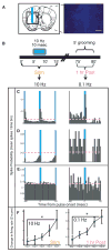
Fig. 3. Repeated daily stimulation of OFC-VMS projections leads to increased evoked firing
(A) (Left) Schematic diagram of stereo-optrode implant site. (Right) Placement visualized via implanting a stereo-optrode dipped in Hoecsht stain (1:1000). Scale bar, 500 μm. (B) Stimulation protocol used for in vivo recording. (C to E) Representative peristimulus spike histograms (5-ms time bins) of three neurons recorded during 10 Hz stimulation (left) and 0.1 Hz probe pulses (1 hour poststimulation on right). Baseline spontaneous firing rate for each cell is shown as pink dashed line. Cells exhibited varied stimulus responsiveness, including evoked activation (C), evoked suppression (D), and no response (E). (F) Light-evoked firing (measured by peristimulus z-scores) across 5 days of stimulation both during 10 Hz stimulation (Stim) and during 0.1 Hz probe pulses 1 hour after stimulation (1 hour post) (*P < 0.021 and P < 0.004). Negative Z-scores for 0.1 Hz on days 1 and 2 indicate net suppression of evoked firing rate during Groom1 hour post after the first two epochs of 10 Hz stimulation.
Fig. 4
Fig. 4. Perseverative grooming and elevated evoked firing rate are resolved by chronic, but not acute, fluoxetine treatment
(A) Experimental time line for fluoxetine wash-out experiment. (B) Two weeks of fluoxetine treatment reduced grooming to level of controls. Main effect: P < 0.009; F = 9.53; Fisher’s PLSD: baseline versus week 2, ***P < 0.003. Increased grooming was reestablished after a 1-week fluoxetine wash-out. Main effect: P < 0.09; F = 3.58. n values: ChR2+ mice = 8; controls = 7. (C) Experimental time line for fluoxetine versus vehicle experiment. (D) Two weeks of fluoxetine treatment reduced grooming to levels of vehicle-treated animals. Main effect: P < 0.14; F = 2.59; Fisher’s PLSD: baseline versus week 2, *P < 0.04. Fluoxetine: n = 7; vehicle: n = 6. (E) (Left) In stereo-optrode–implanted animals, peristimulus Z-scores for 10 Hz stimuli normalized after 2 weeks of fluoxetine (P < 0.028); after 2-week wash-out, Z-scores returned to pretreatment levels. (Right) Peristimulus Z-scores for 0.1 Hz probe pulses showed a nonsignificant decrease after fluoxetine treatment, which returned to pretreatment levels after wash-out.

Comment in

References

    1. Murray CJL, Lopez AD. The Global Burden of Disease: A Comprehensive Assessment of Mortality and Disability from Diseases, Injuries, and Risk Factors in 1990 and Projected to 2020, Global burden of disease and injury series. Vol. 1. Harvard School of Public Health, Harvard Univ Press; Cambridge, MA: 1996.
    1. Kessler RC, et al. Arch Gen Psychiatry. 2005;62:593. - PubMed
    1. Ting JT, Feng G. Curr Opin Neurobiol. 2011;21:842. - PMC - PubMed
    1. Marsh R, Maia TV, Peterson BS. Am J Psychiatry. 2009;166:664. - PMC - PubMed
    1. Milad MR, Rauch SL. Trends Cogn Sci. 2012;16:43. - PMC - PubMed

Publication types

MeSH terms