. 2013 Aug;33(8):1952-9.
doi: 10.1161/ATVBAHA.113.301805. Epub 2013 Jun 6.

Flt-1 (vascular endothelial growth factor receptor-1) is essential for the vascular endothelial growth factor-Notch feedback loop during angiogenesis

Affiliations

Flt-1 (vascular endothelial growth factor receptor-1) is essential for the vascular endothelial growth factor-Notch feedback loop during angiogenesis

John C Chappell et al. Arterioscler Thromb Vasc Biol. 2013 Aug.

Abstract

Objective: Vascular endothelial growth factor (VEGF) signaling induces Notch signaling during angiogenesis. Flt-1/VEGF receptor-1 negatively modulates VEGF signaling. Therefore, we tested the hypothesis that disrupted Flt-1 regulation of VEGF signaling causes Notch pathway defects that contribute to dysmorphogenesis of Flt-1 mutant vessels.

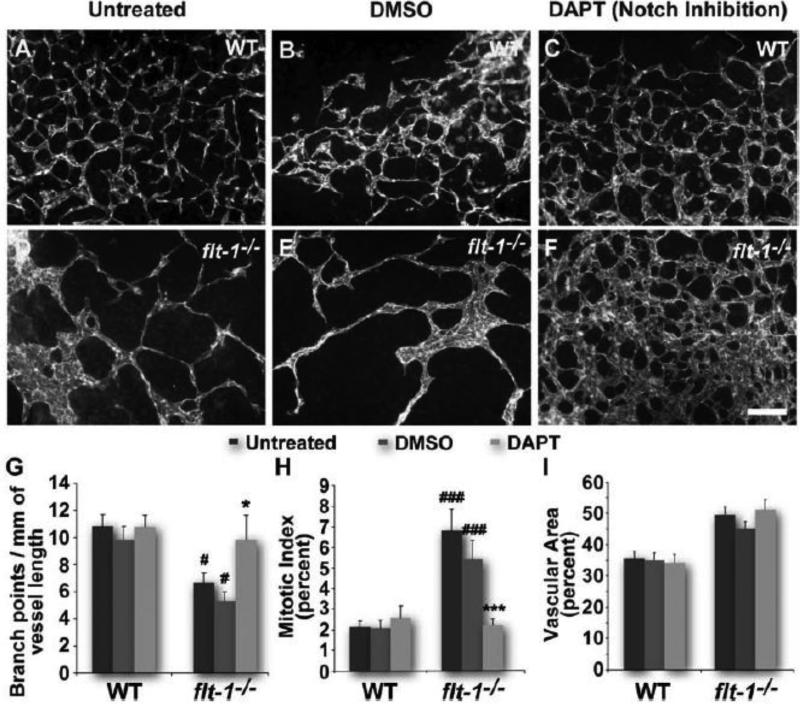
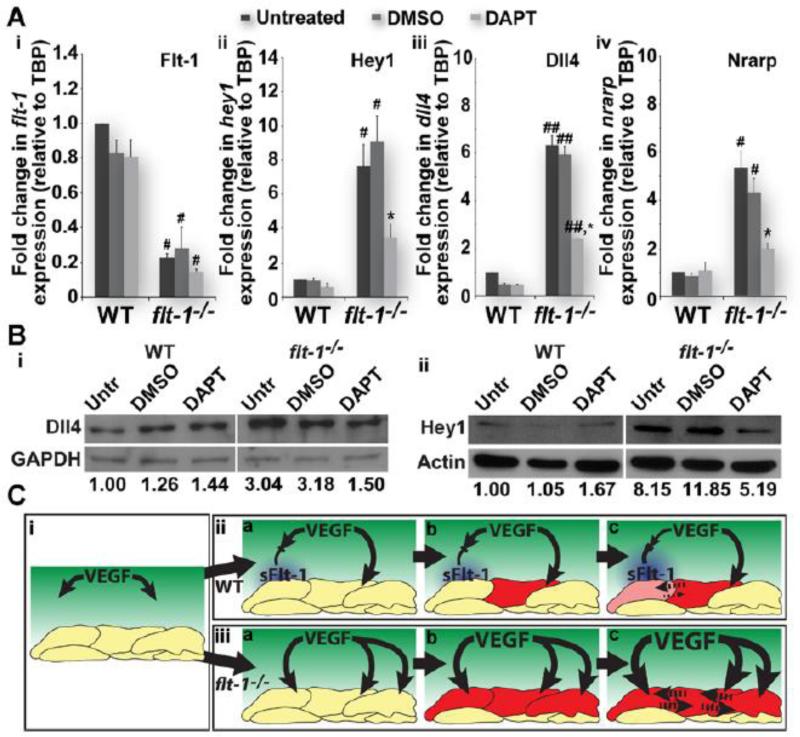
Approach and results: Wild-type and flt-1(-/-) mouse embryonic stem cell-derived vessels were exposed to pharmacological and protein-based Notch inhibitors with and without added VEGF. Vessel morphology, endothelial cell proliferation, and Notch target gene expression levels were assessed. Similar pathway manipulations were performed in developing vessels of zebrafish embryos. Notch inhibition reduced flt-1(-/-) embryonic stem cell-derived vessel branching dysmorphogenesis and endothelial hyperproliferation, and rescue of flt-1(-/-) vessels was accompanied by a reduction in elevated Notch targets. Surprisingly, wild-type vessel morphogenesis and proliferation were unaffected by Notch suppression, Notch targets in wild-type endothelium were unchanged, and Notch suppression perturbed zebrafish intersegmental vessels but not caudal vein plexuses. In contrast, exogenous VEGF caused wild-type embryonic stem cell-derived vessel and zebrafish intersegmental vessel dysmorphogenesis that was rescued by Notch blockade.

Conclusions: Elevated Notch signaling downstream of perturbed VEGF signaling contributes to aberrant flt-1(-/-) blood vessel formation. Notch signaling may be dispensable for blood vessel formation when VEGF signaling is below a critical threshold.

Keywords: Flt-1 protein, mouse; Notch receptors; angiogenesis; embryonic stem cells; vascular endothelial growth factor A; zebrafish.


Figures

Figure 1
Figure 1. Notch inhibition by DAPT rescues the dysmorphogenesis of flt-1−/− blood vessels
Wild-type (A-C) and flt-1−/− (D-F) day 8 ES cell-derived vessels stained for PECAM-1. Scale bar, 100 μm. Dy 8 vessel networks assessed for branch points per vessel length (G). #, p≤0.05 vs. WT of same treatment group. *, p≤0.05 vs. flt-1−/−/untreated or flt-1−/−/DMSO. Dy 7 vessel mitotic indicies were quantified by counting PH3+/PECAM-1+ cells and normalizing to total PECAM-1+ cells (H). ###, p≤0.0005 vs. WT of same treatment group. ***, p≤0.0005 vs. flt-1−/−/untreated or flt-1−/−/DMSO. Vessel area relative to total area for dy 8 ES cell-derived blood vessels (I). Values are averages +/− standard error of the mean (SEM).
Figure 2
Figure 2. Flt-1−/− blood vessel dysmorphogenesis is rescued by Dll4-Fc treatment
Wild-type (A-C) and flt-1−/− mutant (D-F) day 8 ES cell-derived vessels stained for PECAM-1. Scale bar, 100 μm. Dy 8 branch points were counted and normalized to vessel length (G). #, p≤0.05 vs. WT of same treatment group. *, p≤0.05 vs. flt-1−/−/untreated or flt-1−/−/BSA. Mitotic indicies calculated for dy 7 vessels (H). ##, p≤0.005 vs. WT of same treatment group. **, p≤0.005 vs. flt-1−/−/untreated or flt-1−/−/BSA. Dy 8 ES cell-derived vessels assessed for vascular area (I). Values are averages +/− SEM.
Figure 3
Figure 3. Notch inhibition by DAPT disrupts zebrafish intersegmental vessel (ISV) formation but has no effect on the developing caudal vein plexus (CVP)
DMSO-treated (A) and DAPT-treated (B) 48 hpf Tg(kdrl:GFP) zebrafish embryos. Scale bars, 100 μm. Embryos with normal (top inset, A) and defective ISVs (top inset, B), as well as normal (bottom inset, A and B) and defective CVPs, were quantified (C). ###, p≤0.0001 vs. ISV/DMSO. ***, p≤0.0001 vs. ISV/DAPT. Values are percentages.
Figure 4
Figure 4. Notch blockade rescues vessel defects induced by added VEGF
VEGF-treated WT (A-C) and flt-1−/− (D-F) day 8 ES cell-derived vessels stained for PECAM-1. Scale bar, 100 μm. Dy 8 vessels evaluated for branch points per vessel length (G). *, p≤0.05 vs. WT/untreated or WT/VEGF+DAPT. ##, p≤0.002 vs. WT/untreated. ***, p≤0.008 vs. flt-1−/−/untreated, flt-1−/−/VEGF, or flt-1−/−/VEGF+DMSO. Mitotic indicies of dy 7 ES cell-derived vessels (H). *, p≤0.05 vs. WT/VEGF. **, p≤0.01 vs. WT/VEGF or WT/VEGF+DMSO. #, p≤0.05 vs. WT/untreated. ***, p≤0.006 vs. flt-1−/−/untreated, flt-1−/−/VEGF, or flt-1−/−/VEGF+DMSO. Dy 8 vascular area (I). *, p≤0.05 vs. WT/VEGF or WT/VEGF+DMSO. #, p≤0.002 vs WT/untreated. Values are averages +/− SEM.
Figure 5
Figure 5. Notch inhibition by DAPT exacerbates VEGF-A-mediated zebrafish intersegmental vessel (ISV) defects
ISVs from DMSO- and DAPT-treated WT (A-B) and Tg(hsp70l:vegfaa) (C-D) zebrafish embryos at 48 hpf visualized by endothelial expression of GFP [Tg(kdrl:GFP)]. Scale bar, 50 μm. Embryos with affected ISVs (B-D) were quantified, and penetrance was determined as the percent of embryos with an ISV phenotype (E). **, p≤0.005 vs. WT/DMSO. ##, p≤0.007 vs. WT/DMSO. *, p≤0.016 vs. Tg(hsp70l:vegfaa)/DMSO. Values are averages +/− SEM. Of the Tg(hsp70l:vegfaa) embryos with an ISV phenotype, the percent of somites with affected ISVs was determined (F). *, p≤0.0001 for DMSO vs. DAPT. Severities for individual zebrafish are shown as diamonds, with bars representing averages +/− SEM.
Figure 6
Figure 6. Loss of endothelial flt-1 up-regulates the Notch pathway
Flt-1−/− endothelial cell-enriched preps increases Notch target RNAs (A). Real-time quantitative PCR of Flt-1 (Ai) and Notch pathway components Hey1 (Aii), Dll4 (Aiii), and Nrarp (Aiv) from untreated, vehicle control-treated, and DAPT-treated WT and flt-1−/− endothelial cell-enriched preps. (Ai), #, p≤0.05 vs. WT of the same treatment group. (Aii), #, p≤0.05 vs. WT of the same treatment group. *, p≤0.05 vs. flt-1−/−/untreated or flt-1−/−/DMSO. (Aiii), ##, p≤0.008 vs. WT of the same treatment group. *, p≤0.01 vs. flt-1−/−/untreated or flt-1−/−/DMSO. (Aiv), #, p≤0.05 vs. WT of the same treatment group. *, p≤0.05 vs. flt-1−/−/untreated or flt-1−/−/DMSO. Values are averages + SEM. Flt-1−/− endothelial cell-enriched preps have elevated Notch target proteins (B). Representative Western blots for Dll4 (75 kD) and Hey1 (34 kD), as well as GAPDH (36 kD) and actin (45 kD) (for normalization), from untreated, vehicle control-treated, and DAPT-treated WT and flt-1−/− ES cell-derived endothelial cell-enriched preps. Dll4 signal intensities were normalized to those for corresponding GAPDH control bands, and untreated WT levels were set to 1 for comparison (Bi). Hey1 levels were also compared across treatment groups and cell types using actin control bands, just as described for Dll4 and GAPDH (Bii). Model of Flt-1-mediated crosstalk between the VEGF and Notch pathways (C). The model illustrates how Flt-1 (blue), and soluble Flt-1 (sFlt-1) in particular (iia-c), modulates the concentration of available VEGF (green, i-iii) that induces Dll4 expression in endothelial cells (red and pink cells, iia-c). Notch signaling between adjacent cells (dotted lines in iic) then reinforces competition dynamics for sprouting (iic), which completes the Flt-1-mediated feedback loop between VEGF and Notch signaling pathways (iic). In the absence of Flt-1 activity (iiia-c), VEGF induces widespread activation of Dll4 (red cells, iiia-c), and thus Notch signaling is elevated, and normal competition dynamics among endothelial cells are disrupted (dotted lines in iiic). In addition, without Flt-1-mediated feedback, VEGF signaling is unchecked (iiic), exacerbating the excessive Notch signaling and further undermining normal sprouting and proliferation.

References

    1. Potente M, Gerhardt H, Carmeliet P. Basic and therapeutic aspects of angiogenesis. Cell. 2011;146:873–887. - PubMed
    1. Chappell JC, Taylor SM, Ferrara N, Bautch VL. Local guidance of emerging vessel sprouts requires soluble Flt-1. Dev Cell. 2009;17:377–386. - PMC - PubMed
    1. Dorrell MI, Aguilar E, Friedlander M. Retinal vascular development is mediated by endothelial filopodia, a preexisting astrocytic template and specific R-cadherin adhesion. Invest Ophthalmol Vis Sci. 2002;43:3500–3510. - PubMed
    1. Chen TT, Luque A, Lee S, Anderson SM, Segura T, Iruela-Arispe ML. Anchorage of VEGF to the extracellular matrix conveys differential signaling responses to endothelial cells. J Cell Biol. 2010;188:595–609. - PMC - PubMed
    1. Iruela-Arispe ML, Davis GE. Cellular and molecular mechanisms of vascular lumen formation. Dev Cell. 2009;16:222–231. - PMC - PubMed

Publication types

MeSH terms

Substances