. 2013 Jul 2;54(7):4503-11.
doi: 10.1167/iovs.13-12140.

Ablation of the X-linked retinitis pigmentosa 2 (Rp2) gene in mice results in opsin mislocalization and photoreceptor degeneration

Affiliations

Ablation of the X-linked retinitis pigmentosa 2 (Rp2) gene in mice results in opsin mislocalization and photoreceptor degeneration

Linjing Li et al. Invest Ophthalmol Vis Sci. .

Abstract

Purpose: Mutations in the RP2 gene are associated with 10% to 15% of X-linked retinitis pigmentosa (XLRP), a debilitating disorder characterized by the degeneration of retinal rod and cone photoreceptors. The molecular mechanism of pathogenesis of photoreceptor degeneration in XLRP-RP2 has not been elucidated, and no treatment is currently available. This study was undertaken to investigate the pathogenesis of RP2-associated retinal degeneration.

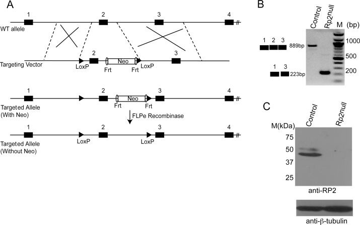
Methods: We introduced loxP sites that flank exon 2, a mutational hotspot in XLRP-RP2, in the mouse Rp2 gene. We then produced Rp2-null allele using transgenic mice that expressed Cre-recombinase under control of the ubiquitous CAG promoter. Electroretinography (ERG), histology, light microscopy, transmission electron microscopy, and immunofluorescence microscopy were performed to ascertain the effect of ablation of Rp2 on photoreceptor development, function, and protein trafficking.

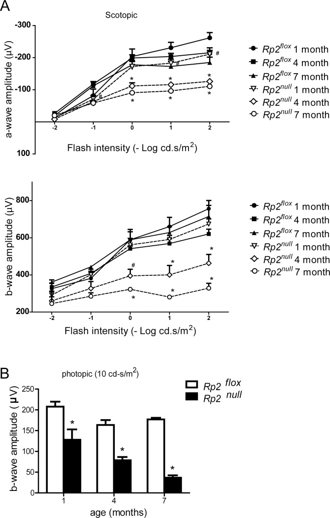
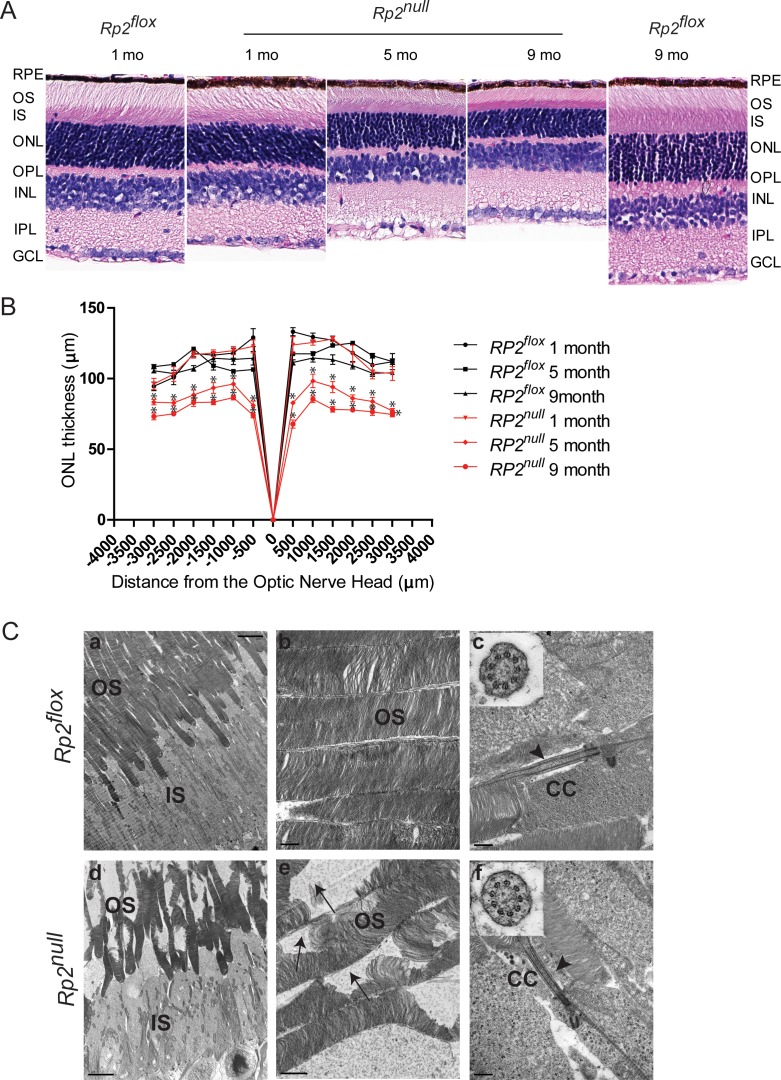
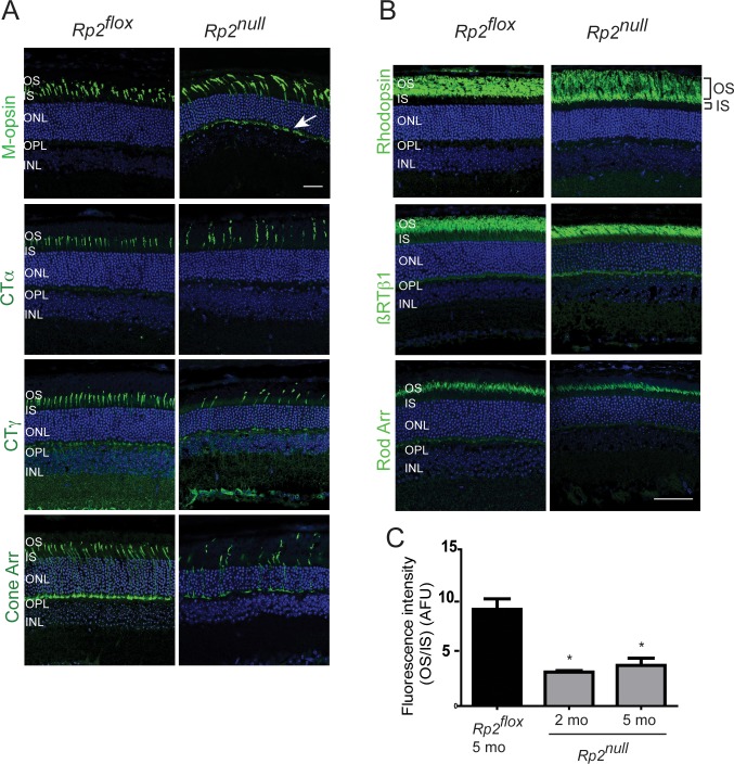
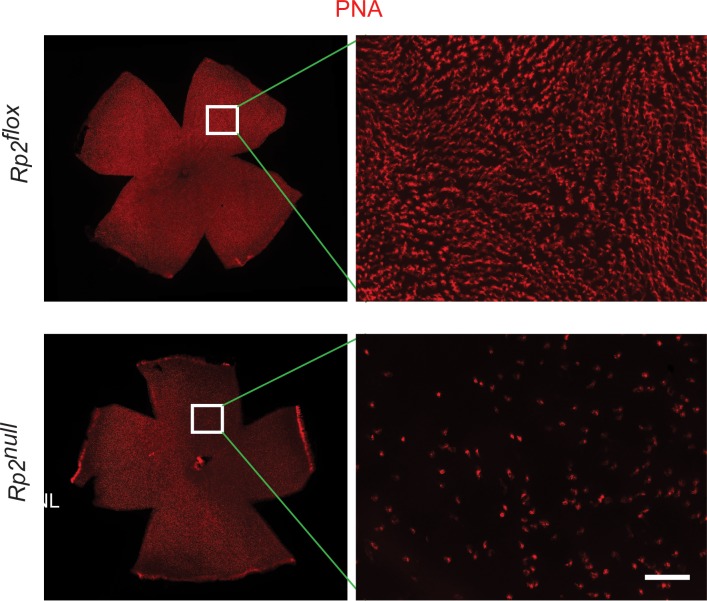
Results: Although no gross abnormalities were detected in the Rp2(null) mice, photopic (cone) and scotopic (rod) function as measured by ERG showed a gradual decline starting as early as 1 month of age. We also detected slow progressive degeneration of the photoreceptor membrane discs in the mutant retina. These defects were associated with mislocalization of cone opsins to the nuclear and synaptic layers and reduced rhodopsin content in the outer segment of mutant retina prior to the onset of photoreceptor degeneration.

Conclusions: Our studies suggest that RP2 contributes to the maintenance of photoreceptor function and that cone opsin mislocalization represents an early step in XLRP caused by RP2 mutations. The Rp2(null) mice should serve as a useful preclinical model for testing gene- and cell-based therapies.

Keywords: Rp2; photoreceptor; retina.


Figures

Figure 1
Figure 1
Generation of conditional allele of Rp2. (A) Schematic of the targeting construct and partial Rp2 genomic DNA showing location of the loxP sites, Frt recombinase, and selectable marker (neomycin [Neo]). The diagram is not drawn to scale. (B) RT-PCR analysis of retinal RNA could detect an expected size band of approximately 889 bp for wild-type (WT) allele in control and a shorter band of approximately 223 bp in Rp2null mice. (C) Immunoblot analysis of retinal protein extracts (50 μg each) using anti-RP2 antibody revealed an expected size band of approximately 40 kDa in control but not in the mutant retina. Immunoblotting using anti-β tubulin antibody (bottom) was used as loading control. Molecular mass marker (M) is shown in kDa.
Figure 2
Figure 2
Photoreceptor dysfunction in Rp2null mice. Rod (scotopic; dark-adapted, [A] and cone (photopic; light-adapted, [B] function were assessed by ERG analysis at indicated ages (n = 5). All data were statistically analyzed using two-tailed Student's t-test and are shown as mean ± SEM. #P < 0.05 and *P < 0.01.
Figure 3
Figure 3
Retinal histology and TEM of Rp2null retina. (A) Histologic analysis of paraffin sections of Rp2flox (control) or Rp2null mouse retina was performed at indicated ages. RPE, retinal pigment epithelium; OS, outer segment; IS, inner segment; ONL, outer nuclear layer; OPL, outer plexiform layer; INL, inner nuclear layer; IPL, inner plexiform layer; GCL, ganglion cell layer. (B) Morphometric analysis of ONL thickness of 1, 5, and 9 months Rp2flox and Rp2null retinas (n = 5). The ONL thickness was measured in μm along the vertical meridian at each defined distance from the optic nerve head. Data represent mean ± SEM. *P < 0.01. (C) TEM analysis of control (Rp2flox, top) and mutant (Rp2null, bottom) photoreceptors at 10 months of age show disorganized OS in the mutant retina. Insets (c, f) show cross-sectional view of the connecting cilium (CC) with 9 + 0 array of microtubules in both control and mutant mice. Arrows in (e) indicate degenerated OS discs in the mutant retina; arrowheads in (c, f) show normal morphology of the CC. Scale: (a, d) 5.0 μm; (b, c, f) 0.5 μm; (e): 1 μm.
Figure 4
Figure 4
Immunofluorescence analysis of Rp2null retinas. Retinal cryosections from 2 (M-opsin) or 5 months (other antibodies) old Rp2flox (control) or Rp2null mice were stained with cone (A) or rod (B) specific proteins, as indicated (green). CTα, cone transducin alpha subunit; CTγ, cone transducin gamma subunit; Cone Arr, cone arrestin; RTβ1, rod transducin beta1 subunit; Rod Arr, rod arrestin. Arrow (in [A]) shows the mislocalization of cone opsin to OPL. (C) Quantitative analysis of rhodopsin signal was performed by analyzing the ratio of fluorescence intensity of rhodopsin 1D4 staining in outer segment to that in the inner segment (OS/IS; the regions are shown in top panel in [B]) of Rp2flox (control) or Rp2null mice. Fluorescence intensity of rhodopsin was at a distance of 1.0 mm from the ONH. The OS/IS ratios from four sections of three mice in each group were analyzed by two-way ANOVA. Means ± SEM (error bars) are shown. *P < 0.05. Data are represented in arbitrary fluorescence units (AFU).
Figure 5
Figure 5
Staining of flat-mount retinas. Flat mount of retinas from Rp2flox (control) or Rp2null mice (10 months) were stained with PNA (red). Inset: The magnified image of the marked area. The mutant retina shows reduction of cones compared with Rp2flox. Scale bar: 50 μm.

References

    1. Kennedy B, Malicki J. What drives cell morphogenesis: a look inside the vertebrate photoreceptor. Dev Dyn. 2009; 238: 2115–2138 - PMC - PubMed
    1. Wright AF, Chakarova CF, Abd El-Aziz MM, Bhattacharya SS. Photoreceptor degeneration: genetic and mechanistic dissection of a complex trait. Nat Rev Genet. 2010; 11: 273–284 - PubMed
    1. Nathans J, Piantanida TP, Eddy RL, Shows TB, Hogness DS. Molecular genetics of inherited variation in human color vision. Science. 1986; 232: 203–210 - PubMed
    1. Neitz M, Neitz J, Jacobs GH. Spectral tuning of pigments underlying red-green color vision. Science. 1991; 252: 971–974 - PubMed
    1. Deretic D. Post-Golgi trafficking of rhodopsin in retinal photoreceptors. Eye. 1998; 12: 526–530 - PubMed

Publication types

MeSH terms