. 2013 Aug 16;8(8):1704-12.
doi: 10.1021/cb300657r. Epub 2013 Jun 19.

A fluorogenic TMP-tag for high signal-to-background intracellular live cell imaging

Affiliations

A fluorogenic TMP-tag for high signal-to-background intracellular live cell imaging

Chaoran Jing et al. ACS Chem Biol. .

Abstract

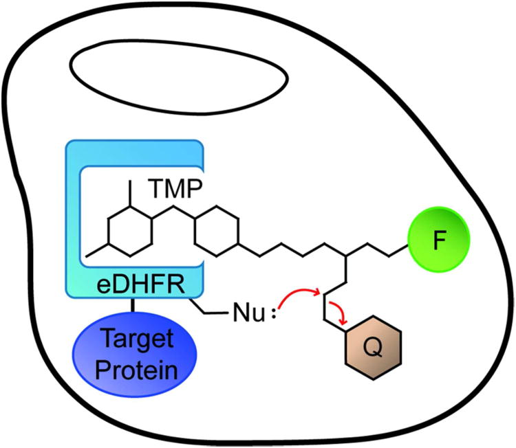
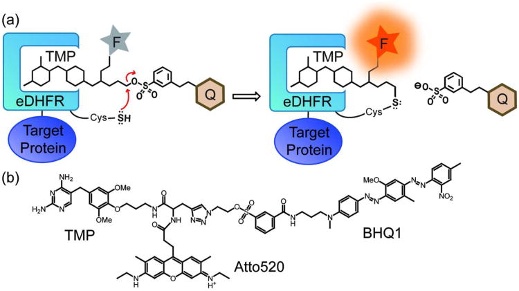
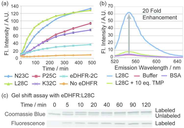
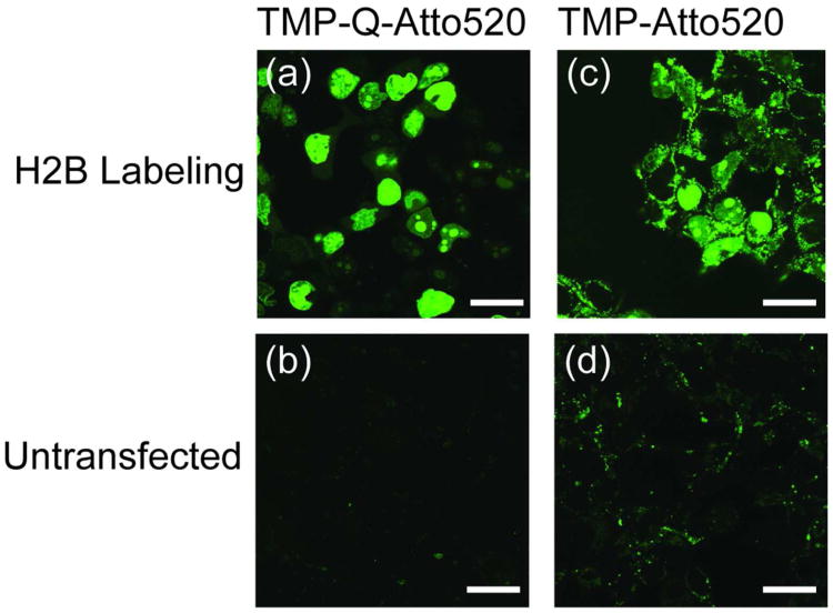
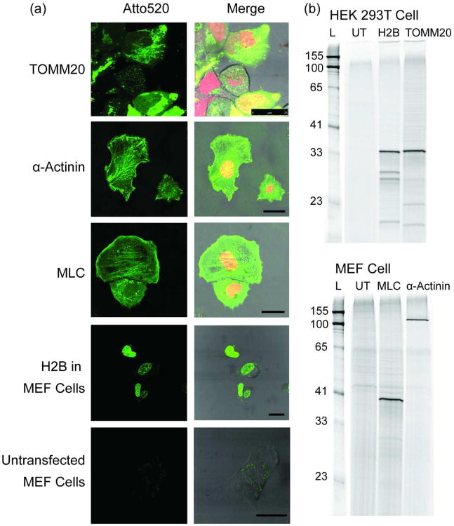
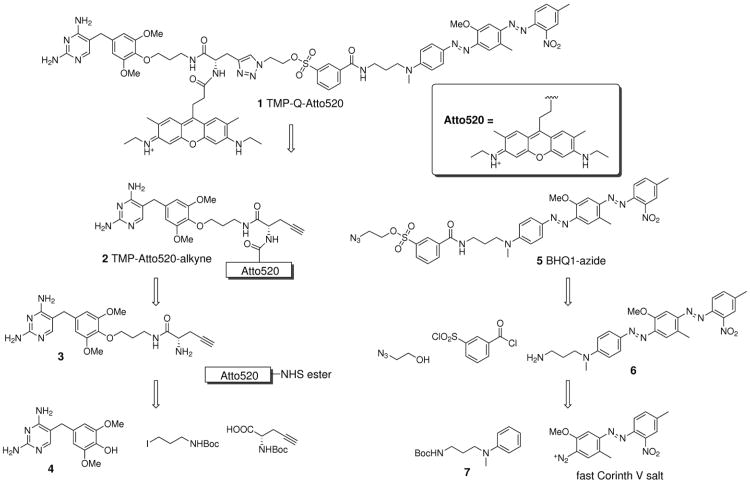
Developed to complement the use of fluorescent proteins in live cell imaging, chemical tags enjoy the benefit of modular incorporation of organic fluorophores, opening the possibility of high photon output and special photophysical properties. However, the theoretical challenge in using chemical tags as opposed to fluorescent proteins for high-resolution imaging is background noise from unbound and/or nonspecifically bound ligand-fluorophore. We envisioned we could overcome this limit by engineering fluorogenic trimethoprim-based chemical tags (TMP-tags) in which the fluorophore is quenched until binding with E. coli dihydrofolate reductase (eDHFR)-tagged protein displaces the quencher. Thus, we began by building a nonfluorogenic, covalent TMP-tag based on a proximity-induced reaction known to achieve rapid and specific labeling both in vitro and inside of living cells. Here we take the final step and render the covalent TMP-tag fluorogenic. In brief, we designed a trimeric TMP-fluorophore-quencher molecule (TMP-Q-Atto520) with the quencher attached to a leaving group that, upon TMP binding to eDHFR, would be cleaved by a cysteine residue (Cys) installed just outside the binding pocket of eDHFR. We present the in vitro experiments showing that the eDHFR:L28C nucleophile cleaves the TMP-Q-Atto520 rapidly and efficiently, resulting in covalent labeling and remarkable fluorescence enhancement. Most significantly, while only our initial design, TMP-Q-Atto520 achieved the demanding goal of not only labeling highly abundant, localized intracellular proteins but also less abundant, more dynamic cytoplasmic proteins. These results suggest that the fluorogenic TMP-tag can significantly impact high-resolution live cell imaging and further establish the potential of proximity-induced reactivity and organic chemistry more broadly as part of the growing toolbox for synthetic biology and cell engineering.


Figures

Figure 1
Figure 1
Cartoon of fluorogenic TMP-tag. Fluorogenic TMP-tag is based on the proximity-induced reaction between the E. coli dihydrofolate reductase (eDHFR) variant and the trimethoprim (TMP) – fluorophore (F) – quencher (Q) heterotrimer. The target protein (purple) is tagged with eDHFR (blue) and then labeled with TMP analogue. The high affinity interaction between TMP and eDHFR induces the SN2 reaction between a nucleophilic amino acid residue (Nu:) engineered on eDHFR and an electrophilic linker attached to the quencher as the leaving group.
Figure 2
Figure 2
The fluorogenic TMP-tag. a) Cartoon of the fluorogenic TMP-tag, which centers a trimeric TMP-quencher (Q) - fluorophore (F) molecule to be cleaved in a proximity-induced SN2 reaction. Specifically, we selected the Cys side chain as the nucleophile and a tosylate linker as the electrophile. When TMP binds to eDHFR, the unique Cys near binding pocket would replace the tosylate leaving group attached to the quencher, and thus the probe is switched on. b) Structure of TMP-BHQ1-Atto520 (TMP-Q-Atto520), the first fluorogenic TMP-tag.
Figure 3
Figure 3
Depiction of the designed eDHFR:Cys mutant library. Cartoon of TMP bound to eDHFR, with all 16 amino acid residues around the binding pocket mutated to Cys highlighted. The eDHFR protein is represented in violet as a ribbon diagram, with the highlighted residues in orange. The TMP is represented as sticks with coloring based on elements. Since no structure has been solved of TMP bound to eDHFR, the model was created by structural alignment of eDHFR with the L. casei DHFR. The graphic was prepared using PyMOL.
Figure 4
Figure 4
In vitro reactivity of fluorogenic TMP-tag. To demonstrate the specific fluorescence enhancement and efficiency of covalent labeling, TMP-Q-Atto520 was characterized with purified eDHFR:Cys variants in fluorometry and gel-shift assays. a) Screening of the eDHFR:Cys variant library. Purified eDHFR:Cys variants at a concentration of 1 μM were incubated with 1 μM TMP-Q-Atto520 in PBS (pH = 7.40) with 100 μM NADPH and 1 mM glutathione at 37 °C. Fluorescence intensity (Fl. Intensity) was measured at various time points. Four eDHFR:Cys mutants, namely the eDHFR:N23C, P25C, L28C and K32C showed significant fluorescence enhancement. As controls, the eDHFR-2C variant that lacks Cys and the buffer without eDHFR induced little increase in fluorescence intensity. Error bars represent standard deviation. b) The fluorescence emission spectra of TMP-Q-Atto520. Purified eDHFR:L28C was labeled under the same reaction conditions as in (a) for 3 h. The fluorescence spectrum showed 20 fold enhancement compared to the control group in which 1 μM TMP-Q-Atto520 was incubated with the buffer without eDHFR:L28C. By contrast, 1 μM TMP-Q-Atto520 incubated with 1 μM bovine serum albumin (BSA) which does not bind TMP shows no significant fluorescence enhancement compared to control group with buffer. In addition, the specific quencher cleavage is further demonstrated by the competition assay in which 1 μM TMP-Q-Atto520 was incubated with 1 μM eDHFR:L28C and 10 μM TMP. Standard deviation < 5% and error bards are not shown. c) Determination of the rate of covalent labeling. In the same buffer as in (a), 2 μM of eDHFR:L28C was incubated with 10 μM TMP-Q-Atto520 at 37 °C. At various time points, aliquots were quenched by boiling at 95 °C with 6X SDS. The reaction was analyzed by sodium dodecyl sulfate (SDS)-PAGE followed by fluorescence gel scanning and Coomassie staining, and it was determined that 50% labeling occurs in approximately 10 minutes.
Figure 5
Figure 5
Live cell imaging of cell nucleus with fluorogenic TMP-tag in comparison with non-fluorogenic TMP-tag. a) HEK 293T cells were transiently transfected with plasmid encoding histone 2B (H2B)-eDHFR:L28C and labeled with 5 μM TMP-Q-Atto520 for 3 h. Live cell imaging was achieved using confocal microscope, displaying the specific labeling of cell nucleus. b) As control, untransfected cells were treated with TMP-Q-Atto520 under the same condition as (a), showing very weak background fluorescence. c, d) The same imaging experiments were carried out with the first generation, noncovalent TMP-tag. Significant background staining was observed with both H2B-eDHFR transfected and untransfected cells. For comparison, all four images were obtained using the same microscope setup with excitation at 488 nm. Scale bars are 25 μm.
Figure 6
Figure 6
Live cell imaging of cytosolic proteins with TMP-Q-Atto520. Three cytosolic proteins, TOMM20, α-actinin, MLC, in addition to H2B were successfully imaged in two distinct cell lines. a) Live cell imaging using TMP-Q-Atto520. HEK 293T cells (for TOMM20) or mouse embryonic fibroblast (MEF) cells (for MLC, α-actinin, and H2B) transiently co-transfected with vectors encoding target protein-eDHFR:L28C and H2B-mCherry (for TOMM20, MLC and α-actinin labeling) fusion proteins, respectively, were incubated with 10 μM TMP-Q-Atto520 in media for 3 h at 37 °C, washed twice with media, and imaged using confocal and differential interference contrast (DIC) microscopy. Untransfected MEF cells were treated under the same condition and imaged using the same microscope setup as for H2B in MEF cells. Left column shows fluorescence imaging from the green channel (488 nm excitation); right column shows merges images from the green, red (594 nm excitation), and DIC channels. Scale bars are 25 μm. b) In-gel fluorescence analysis to confirm the labeling specificity. The cells transfected with corresponding target protein-eDHFR:L28C vectors were harvested after 3 h incubation with 10 μM TMP-Q-Atto520, lysed, and analyzed by SDS-PAGE and in-gel fluorescence scanning with an excitation laser at 488 nm. As controls, untransfected (UT) cells were stained with TMP-Q-Atto520 under the same condition and analyzed in parallel. These results show the target versatility of the fluorogenic TMP-tag for live cell protein labeling.
Scheme 1
Scheme 1
Retro-synthetic design of TMP-Q-Atto520.

References

    1. Chalfie M, Tu Y, Euskirchen G, Ward WW, Prasher DC. Green fluorescent protein as a marker for gene expression. Science. 1994;263:802–805. - PubMed
    1. Heim R, Prasher DC, Tsien RY. Wavelength mutations and posttranslational autoxidation of green fluorescent protein. Proc Natl Acad Sci U S A. 1994;91:12501–12504. - PMC - PubMed
    1. Tsien RY. The green fluorescent protein. Annu Rev Biochem. 1998;67:509–544. - PubMed
    1. Ormo M, Cubitt AB, Kallio K, Gross LA, Tsien RY, Remington SJ. Crystal structure of the Aequorea victoria green fluorescent protein. Science. 1996;273:1392–1395. - PubMed
    1. Shaner NC, Patterson GH, Davidson MW. Advances in fluorescent protein technology. J Cell Sci. 2007;120:4247–4260. - PubMed

Publication types

LinkOut - more resources