. 2013 Jun 6;50(5):661-74.
doi: 10.1016/j.molcel.2013.05.011.

Kinetochores coordinate pericentromeric cohesion and early DNA replication by Cdc7-Dbf4 kinase recruitment

Affiliations

Kinetochores coordinate pericentromeric cohesion and early DNA replication by Cdc7-Dbf4 kinase recruitment

Toyoaki Natsume et al. Mol Cell. .

Abstract

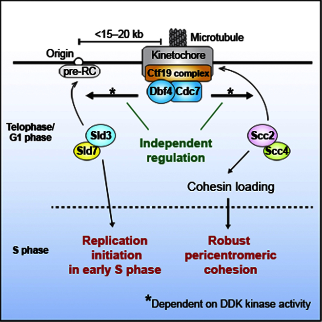
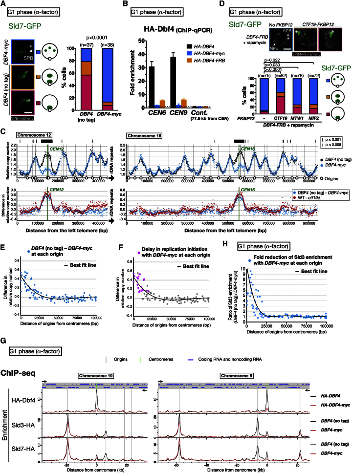
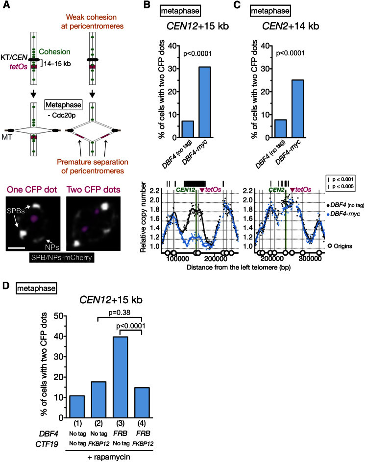
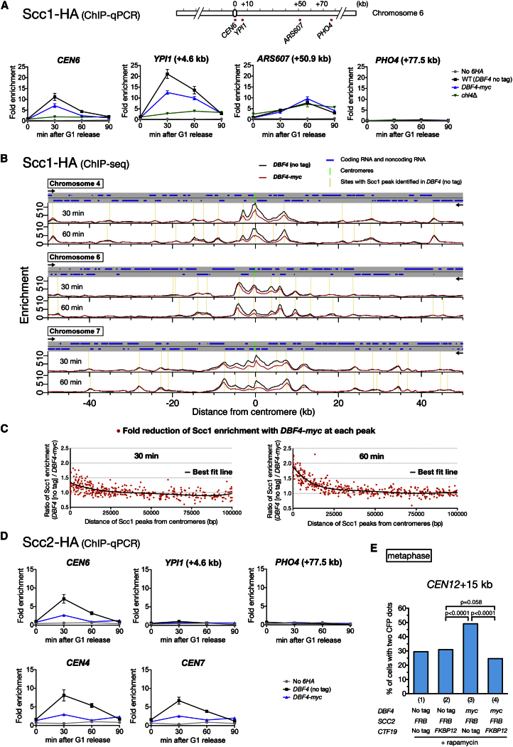
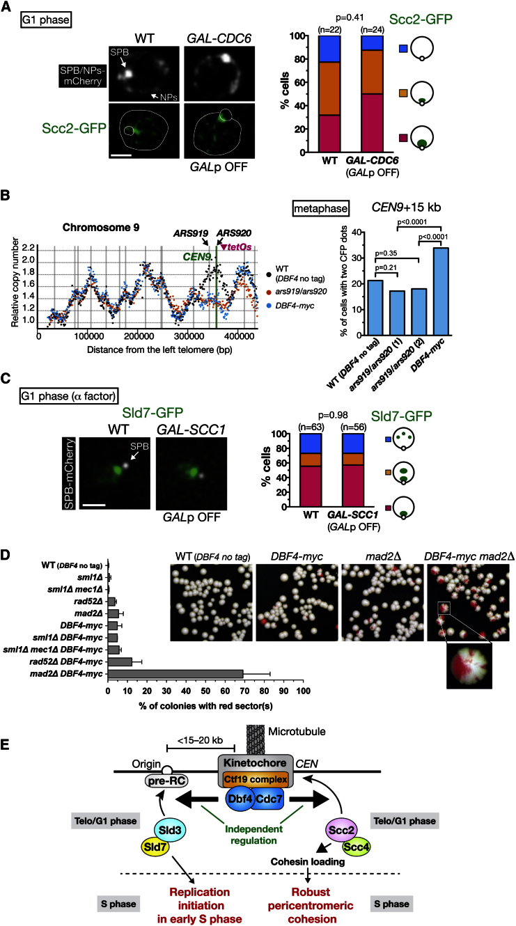
Centromeres play several important roles in ensuring proper chromosome segregation. Not only do they promote kinetochore assembly for microtubule attachment, but they also support robust sister chromatid cohesion at pericentromeres and facilitate replication of centromeric DNA early in S phase. However, it is still elusive how centromeres orchestrate all these functions at the same site. Here, we show that the budding yeast Dbf4-dependent kinase (DDK) accumulates at kinetochores in telophase, facilitated by the Ctf19 kinetochore complex. This promptly recruits Sld3-Sld7 replication initiator proteins to pericentromeric replication origins so that they initiate replication early in S phase. Furthermore, DDK at kinetochores independently recruits the Scc2-Scc4 cohesin loader to centromeres in G1 phase. This enhances cohesin loading and facilitates robust pericentromeric cohesion in S phase. Thus, we have found the central mechanism by which kinetochores orchestrate early S phase DNA replication and robust sister chromatid cohesion at microtubule attachment sites.


Figures

None
Graphical abstract
Figure 1
Figure 1
Replication Factories Are Initially Formed at Centromeric Regions (A) Replication factories are initially formed near the SPB. GFP-PCNA (T8375 [i]) and POL1-GFP (T7300 [ii]) cells with SPC42-mCherry NIC96-mCherry (visualizing SPBs and nuclear pores [NPs]) were observed in asynchronous culture. T7300 cells also had NET1-mCherry (nucleolus marker). We set 0 s arbitrarily. The scale bar represents 1 μm. (B) Replication factories are initially formed, overlapping with the kinetochore cluster. Cells (T10459) with GFP-PCNA SPC42-mCherry NIC96-mCherry NDC80-CFP (visualizing kinetochores [KTs]) were observed in asynchronous culture. White lines represent the nucleus and the SPB. CFP, cyan fluorescent protein. (C) Replication factories are initially formed on centromeres when they are detached from a spindle pole. Cells (T8819) with GFP-PCNA MTW1-CFP NDC80-CFP (KTs-CFP) SPC42-mCherry NIC96-mCherry mad2Δ bub2Δ were arrested with α factor and released into medium with nocodazole. Kinetochores were frequently detached from a spindle pole after going through S phase in the presence of nocodazole. mad2Δ bub2Δ allowed cells to exit from mitosis in the absence of microtubules. Cells were observed after cytokinesis (125 min after release from α factor).
Figure 2
Figure 2
DDK and Sld3–Sld7 Localize at Centromeric Regions from Telophase to G1 Phase (A) DDK and Sld3–Sld7 localize at centromeric regions from telophase to G1 phase. Cells with CDC7-GFP MTW1-CFP NDC80-CFP SPC42-mCherry NIC96-mCherry (T10876 [i]), GFP-DBF4 MTW1-CFP NDC80-CFP (T9466 [ii]), SLD7-GFP NDC80-CFP SPC42-mCherry NIC96-mCherry (T10875 [iii]), and SLD3-GFP SPC42-mCherry (T8784 [iv]) were observed in asynchronous culture. Cells in telophase–G1 phase were selected. The scale bar represents 1 μm. (B) Cdc7 and Sld7 colocalize with centromeres when they are detached from a spindle pole. T8888 and T8886 cells (same as T10876 and T10875 but with mad2Δ bub2Δ) were observed as in Figure 1C. (C) Sld7 localization at centromeric regions is dependent on pre-RC. CDC6+ (wild-type [WT], T9126) and GAL-CDC6 (T9127) cells with SLD7-GFP SPC42-CFP (i), as well as ORC2+ (WT, T9273) and GAL-orc2-1 (T9272) cells with SLD7-GFP SPC42-mCherry NIC96-mCherry rad9Δ rad24Δ mad2Δ (ii), were treated with α factor, released from it, and treated with α factor again. GAL promoter (GALp) was shut off during this process (see Supplemental Experimental Procedures). Cells were observed after cytokinesis. (D) Cdc7 localization on centromeric regions is independent of pre-RC. GAL-CDC6 cells (T9125) with CDC7-GFP SPC42-CFP NIC96-mCherry (i), as well as ORC2+ (WT, T9275) and GAL-orc2-1 (T9274) cells with CDC7-GFP SPC42-mCherry NIC96-mCherry rad9Δ rad24Δ mad2Δ (ii), were treated with α factor, released from it, and observed in 120 min. GALp was active (ON) or shut off (OFF). (E) Cdc7 and its kinase activity are required for Sld7 localization on centromeric regions. (i) CDC7+ (WT, T8613) and cdc7-4 (T9085) cells with SLD7-GFP SPC42-mCherry were incubated at 25°C for 2.5 hr and then at 37°C for 35 min, in the presence of α factor. (ii) CDC7+ (WT, T8613) and cdc7-as3 (T10417) cells with SLD7-GFP SPC42-mCherry were arrested in G1 phase with α factor and then treated with 20 μM PP1 (inhibitor) for 20 min in the presence of α factor. (F) Summary of results in Figure 2. See details in the text. See also Figure S1.
Figure 3
Figure 3
The Ctf19 Complex Recruits DDK to Centromeres and Facilitates Early Replication of Centromeric Regions (A) Sld7 localization on centromeric regions is abolished in mutants of the Ctf19-complex components. WT (T8613), ctf19Δ (T9650), ctf3Δ (T9708), chl4Δ (T9709), and mcm21Δ (T9710) cells with SLD7-GFP SPC42-mCherry were observed in asynchronous culture. Cells in telophase–G1 phase were selected. The scale bar represents 1 μm. (B) Dbf4 association with centromeres is reduced in ctf19Δ and chl4Δ cells. WT (T9945), ctf19Δ (T10275), and chl4Δ (T10278) cells with HA-DBF4 were treated with α factor for 2.5 hr and processed for ChIP using a hemagglutinin (HA) antibody. Coprecipitated DNA was analyzed with qPCR at CEN6, CEN9, and a control locus (PHO4, 77.5 kb from CEN6). The ratio of immunoprecipitated DNA to total DNA in whole-cell extract is normalized relative to a control locus in WT (fold enrichment). Error bars represent SD. (C) Dbf4 association with replication origins in G1 phase. ChIP-qPCR was performed as in (B) and analyzed at replication origins and the nonorigin (nonORI) locus (PHO4). Error bars represent SD. (D) Replication of centromeric regions is specifically delayed in ctf19Δ cells. S phase and G2–M phase cells were collected from a culture of WT (T9475) and ctf19Δ (T10117) homozygous diploids. The ratio of the copy number in S phase cells to that in G2–M phase cells is normalized and shown between 1.0 and 2.0 at each chromosome locus. The difference in the replication timing between the two strains is shown at bottom. Smoothed lines were added in both graphs. See also Figures S2 and S3.
Figure 4
Figure 4
DDK at Kinetochores Advances Replication of Centromeric Regions in S Phase (A) Sld7 localization is abolished at centromeric regions in G1 phase when Dbf4 is tagged at its C terminus. DBF4 (no tag) (T8613) and DBF4-myc (T9396) cells with SLD7-GFP SPC42-mCherry were treated with α factor for 2.5 hr. The scale bar represents 1 μm. (B) Dbf4 association with centromeres is reduced in G1 phase when Dbf4 is tagged with Myc or FRB at its C terminus. HA-DBF4 (T9945), HA-DBF4-myc (T9979), and HA-DBF4-FRB (T10360) cells were analyzed as in Figure 3B. Error bars represent SD. (C) Replication of centromeric regions is specifically delayed in DBF4-myc cells similarly to ctf19Δ cells. DBF4 (no tag) (T9475) and DBF4-myc (T9476) homozygous diploid cells were analyzed as in Figure 3D. Replication timing (top) and its difference (bottom) are shown. (D) Artificial tethering of Dbf4-FRB to Ctf19-FKBP12 restores Sld7 localization on centromeric regions in G1 phase. DBF4-FRB SLD7-GFP SPC42-mCherry NIC96-mCherry cells with either CTF19-FKBP12 (T10149), MTW1-FKBP12 (T10147), MIF2-FKBP12 (T10148), or no FKBP12 tag (T9915), were treated with α factor for 2.5 hr in the presence of 10 μM rapamycin. (E) Replication timing of origins in DBF4 (no tag) and DBF4-myc cells. Difference in replication timing between in DBF4 (no tag) and DBF4-myc cells (see C) is plotted for origins against the distance from the centromere. A regression curve is shown as a black line. (F) Pericentromeric origins up to 15–20 kb from centromeres show delays in their replication initiation in DBF4-myc cells. Replication origins showing the delay in replication initiation, obtained with mathematical modeling, are marked in magenta in the graph presented in (E). (G) ChIP-seq analysis of Dbf4 and Sld3–Sld7 localization in G1 phase. HA-DBF4 (T9945), HA-DBF4-myc (T9979), SLD3-HA DBF4 (no tag) (T9861), SLD3-HA DBF4-myc (T9970), SLD7-HA DBF4 (no tag) (T9862), and SLD7-HA DBF4-myc (T9971) cells were treated with α factor for 2.5 hr and analyzed via ChIP-seq (immunoprecipitation [IP] with an HA antibody). (H) Pericentromeric origins up to 15–20 kb from centromeres show reduction in their association with Sld3 in DBF4-myc cells. The ratio of Sld3 enrichment (DBF4 [no tag]/DBF4-myc), obtained with ChIP-seq (Figure 4G), is plotted for origins against the distance from the centromere. A regression curve is shown as a black line. See also Figures S3 and S4.
Figure 5
Figure 5
Kinetochore-Associated DDK Facilitates Robust Sister Chromatid Cohesion at Pericentromeres (A) An assay for evaluating cohesion at pericentromeres. The tetOs, integrated at 14–15 kb from a centromere, were bound with TetR-CFP. Cells with CDC20 under MET3 promoter were treated with α factor, released to methionine-containing medium (for depletion of Cdc20), and arrested in metaphase. At 2 hr after the release, the percentage of cells with two sister CFP dots was counted. The scale bar represents 1 μm. (B and C) Sister chromatid cohesion is weakened at the CEN12 and CEN2 pericentromeres in DBF4-myc cells. DBF4 (no tag) (T10141, n = 249) and DBF4-myc (T10142, n = 266) cells with tetOs at +15 kb from CEN12 (B), or those (T10194, n = 347; T10195, n = 299; respectively) with tetOs at +14 kb from CEN2 (C), were treated and analyzed as in (A). The replication-timing profile of the pericentromeres is shown at the bottom. (D) Artificial tethering of Dbf4-FRB to Ctf19-FKBP12 restores pericentromeric cohesion. Cells with indicated alleles of DBF4 and CTF19, as well as tetOs at +15 kb from CEN12, were treated and analyzed as in (A). During α factor treatment and thereafter, 10 μM rapamycin was added. n = 305–353 in each condition. See also Figure S5.
Figure 6
Figure 6
DDK at Kinetochores Facilitates Recruitment of Cohesin Loader and Enriches Cohesins at Pericentromeres (A) Cohesin Scc1 is reduced at a centromere (CEN6) and a pericentromere (YPI1), but not at a chromosome-arm site (ARS607) in DBF4-myc cells. SCC1-HA cells with WT (DBF4 no tag) (T9882), DBF4-myc (T10007), and chl4Δ (T10561) were treated with α factor, released to fresh medium, aliquoted at indicated times, and processed for ChIP-qPCR (IP with an HA antibody). Cells lacking HA tags (T7107) were included as a control. Fold enrichment was as in Figure 3B (CEN6 in WT at 0 min is set to 1). Error bars represent SD. (B) ChIP-seq analysis of Scc1 localization. T9882 and T10007 cells (see A) were treated as in (A) and analyzed by ChIP-seq (IP with an HA antibody). Scc1 peaks were identified using a peak-finding algorithm in cells with DBF4 (no tag) at each time point. (C) Scc1 localization is specifically reduced up to 20 kb from centromeres. The ratio of Scc1 enrichment (DBF4 [no tag]/DBF4-myc) at each peak identified in (B) is plotted against the distance from the centromere. The black line represents a regression curve. (D) The association of cohesin loader Scc2 with the centromere is reduced in DBF4-myc cells. DBF4 (no tag) (T9883) and DBF4-myc (T10008) cells with SCC2-HA were analyzed as in (A). Error bars represent SD. (E) Artificial tethering of Scc2-FRB to Ctf19-FKBP12 strengthens pericentromeric cohesion when DDK is reduced at kinetochores. Cells with indicated alleles of DBF4, SCC2, and CTF19, as well as tetOs at +15 kb from CEN12, were treated and analyzed as in Figure 5D. n = 365–428 in each condition. See also Figure S6.
Figure 7
Figure 7
DDK at Kinetochores Independently Regulates Replication Timing and Cohesion at Pericentromeres (A) Scc2 accumulation at centromeric regions does not require the pre-RC in G1 phase. CDC6+ (WT, T10558) and GAL-CDC6 (T10559) cells with SCC2-GFP SPC42-mCherry NIC96-mCherry were treated as in Figure 2Di. The scale bar represents 1 μm. (B) Inactivation of CEN9-proximal origins does not weaken cohesion at the CEN9 pericentromere. WT (DBF4 no tag, T10461, n = 282), ars919/ars920 (clone 1, T10552, n = 314; clone 2, T10770, n = 316), and DBF4-myc (T10528, n = 295) cells with tetOs at +15 kb from CEN9 were treated as in Figure 5A and analyzed at 160 min after release from α factor. Left: replication timing of chromosome 9. (C) Cohesin is not required for Sld7 accumulation at centromeric regions in G1 phase. SCC1+ (WT, T9110) and GAL-SCC1 (T9107) cells with SLD7-GFP SPC42-mCherry were analyzed as in Figure 2Ci. (D) DBF4-myc cells show an increase in chromosome loss (p = 0.0006), which is further enhanced by deletion of MAD2 (p < 0.0001). Indicated strains with the CFIII chromosome fragment were used for a chromosome loss assay. Loss of CFIII generated red sector in colonies. Error bars represent SD. (E) Summary of two functions of DDK recruited to kinetochores. See details in the text. See also Figure S7.

References

    1. Ahmad K., Henikoff S. Centromeres are specialized replication domains in heterochromatin. J. Cell Biol. 2001;153:101–110. - PMC - PubMed
    1. Akiyoshi B., Nelson C.R., Ranish J.A., Biggins S. Quantitative proteomic analysis of purified yeast kinetochores identifies a PP1 regulatory subunit. Genes Dev. 2009;23:2887–2899. - PMC - PubMed
    1. Aparicio O.M. Location, location, location: it’s all in the timing for replication origins. Genes Dev. 2013;27:117–128. - PMC - PubMed
    1. Araki H. Initiation of chromosomal DNA replication in eukaryotic cells; contribution of yeast genetics to the elucidation. Genes Genet. Syst. 2011;86:141–149. - PubMed
    1. Bailis J.M., Bernard P., Antonelli R., Allshire R.C., Forsburg S.L. Hsk1-Dfp1 is required for heterochromatin-mediated cohesion at centromeres. Nat. Cell Biol. 2003;5:1111–1116. - PubMed

Publication types

MeSH terms

Associated data