. 2013 Jun 4;104(11):2410-8.
doi: 10.1016/j.bpj.2013.04.039.

Small-molecule photostabilizing agents are modifiers of lipid bilayer properties

Affiliations

Small-molecule photostabilizing agents are modifiers of lipid bilayer properties

Jose L Alejo et al. Biophys J. .

Abstract

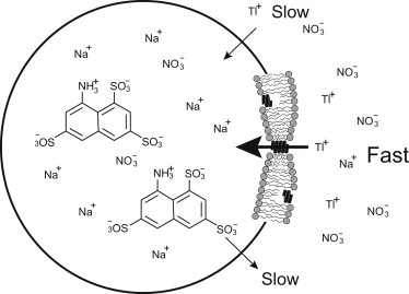
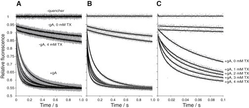
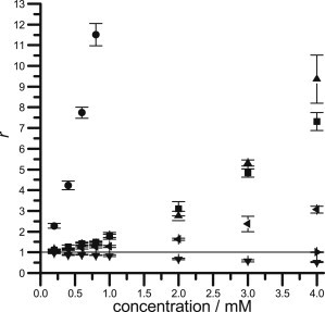
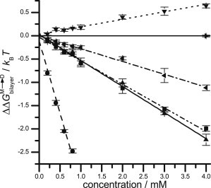
Small-molecule photostabilizing or protective agents (PAs) provide essential support for the stability demands on fluorescent dyes in single-molecule spectroscopy and fluorescence microscopy. These agents are employed also in studies of cell membranes and model systems mimicking lipid bilayer environments, but there is little information about their possible effects on membrane structure and physical properties. Given the impact of amphipathic small molecules on bilayer properties such as elasticity and intrinsic curvature, we investigated the effects of six commonly used PAs--cyclooctatetraene (COT), para-nitrobenzyl alcohol (NBA), Trolox (TX), 1,4-diazabicyclo[2.2.2]octane (DABCO), para-nitrobenzoic acid (pNBA), and n-propyl gallate (nPG)--on bilayer properties using a gramicidin A (gA)-based fluorescence quench assay to probe for PA-induced changes in the gramicidin monomer↔dimer equilibrium. The experiments were done using fluorophore-loaded large unilamellar vesicles that had been doped with gA, and changes in the gA monomer↔dimer equilibrium were assayed using a gA channel-permeable fluorescence quencher (Tl⁺). Changes in bilayer properties caused by, e.g., PA adsorption at the bilayer/solution interface that alter the equilibrium constant for gA channel formation, and thus the number of conducting gA channels in the large unilamellar vesicle membrane, will be detectable as changes in the rate of Tl⁺ influx-the fluorescence quench rate. Over the experimentally relevant millimolar concentration range, TX, NBA, and pNBA, caused comparable increases in gA channel activity. COT, also in the millimolar range, caused a slight decrease in gA channel activity. nPG increased channel activity at submillimolar concentrations. DABCO did not alter gA activity. Five of the six tested PAs thus alter lipid bilayer properties at experimentally relevant concentrations, which becomes important for the design and analysis of fluorescence studies in cells and model membrane systems. We therefore tested combinations of COT, NBA, and TX; the combinations altered the fluorescence quench rate less than would be predicted assuming their effects on bilayer properties were additive. The combination of equimolar concentrations of COT and NBA caused minimal changes in the fluorescence quench rate.


Figures

Figure 1
Figure 1
Using gA channels as probes for changes in lipid bilayer properties. Bilayer-spanning gA channels form by transmembrane dimerization of two β6.3-helical subunits, with a hydrophobic length that is less than the hydrophobic length of the unperturbed bilayer. (Left) Because the channel length (l) is less than the unperturbed bilayer thickness (d0), there is a hydrophobic mismatch; the bilayer will adapt to minimize the exposure of hydrophobic residues to water. This adaptation involves local compression of the bilayer hydrophobic core and bending of the bilayer/solution interface. Channel formation thus will incur an energetic cost, meaning that the energetic cost of channel formation varies with changes in lipid bilayer properties. (Right) In bilayers that have been modified by the adsorption of amphiphiles at the bilayer/solution interface, the bilayer has additional degrees of freedom in terms of how it adapts to a channel-bilayer hydrophobic mismatch because the amphiphiles may redistribute between the membrane and the aqueous phases, which will tend to reduce the magnitude of the energetic cost of channel formation such that the gA monomer↔dimer equilibrium will tend to be shifted toward the conducting dimers. (In some cases, e.g., our results with COT, the compound increases the energetic cost of channel formation and shifts the gA monomer↔dimer equilibrium toward the nonconducting monomers.)
Figure 2
Figure 2
Testing for bilayer-modifying effects using stopped-flow spectrofluorometry. ANTS-loaded LUVs formed with gramicidin A (gA) are exposed to the quencher Tl+. Over the experiment timescale of 1 s, ANTS and TlNO3 are only slightly permeant through the lipid bilayer, whereas gA channels are highly Tl+ permeable. The rate of Tl+ influx is proportional to the number of conducting channels in the LUV membrane. (Expanded view on the right) Segment of the lipid bilayer with the two major gA conformers: nonconducting monomers and conducting dimers. Modified after (40).
Figure 3
Figure 3
Effect of Trolox (TX) on the time course of ANTS fluorescence quenching. (A) (Traces) Normalized fluorescence time signal over 1 s; (shaded dots) results from all repeats (n > 5 per condition); (solid lines) average of all repeats. (Top trace) Results in the absence of the quencher Tl+; (next two traces) results in the absence of gA with Tl+ and without or with the highest TX concentration employed (4 mM); (lower five fluorescence time courses, from top to bottom) gA-containing vesicles incubated with 0, 1, 2, 3, and 4 mM TX. (B) (Dots) Results from a single repeat for each condition; (lines) modified stretched exponential fits to those repeats. (C) The first 100 ms of the fluorescence time courses and their corresponding fits.
Figure 4
Figure 4
Normalized gA activation-induced fluorescence quenching rates (r = 〈k〉/〈kref); see Eq. 7, quantifying the effect of photostabilizing agents on lipid bilayer properties. The symbols denote: ▾, COT; ▸, DABCO; ▴, NBA; ◂, pNBA; ●, nPG; and ▪, TX. (Straight line at r = 1) Guide to the eye.
Figure 5
Figure 5
Shifts in the bilayer contribution to the energetic cost of gA dimer formation due to the influence of various PAs at the specified concentrations. The symbols denote: ▾, COT; ▸, DABCO; ▴, NBA; ◂, pNBA; ●, nPG; and ▪, TX. (Dotted line) Linear fits to the data for COT (y = 0.160x + 0.03, r2 = 0.982); (dot-dashed line) for pNBA (y = –0.275x – 0.005, r2 = 0.994); (dot-dot-dashed line) for TX (y = –0.505x – 0.033, r2 = 0.995); (solid line) for NBA (y = –0.558x + 0.026, r2 = 0.996); and (dashed line) for nPG (slope = –3.096x – 0.1134, r2 = 0.988).
Figure 6
Figure 6
Shifts in the bilayer contribution to the energetic cost of gA dimer formation due to the influence of various PA combinations at the specified concentrations. The symbols denote: ▪, COT+NBA+TX; ●, COT+NBA; ▾, COT+TX; and ▴, NBA+TX. The concentrations indicated on the abscissa denote the concentration of each of the PAs, which were added as equimolar mixtures. (Straight line at ΔΔGbilayer = 0) Guide to the eye. (Dotted line) Linear fits for COT+NBA+TX (y = –0.578x + 0.028, r2 = 0.91); (dot-dashed line) NBA+TX (y = –0.776x – 0.02, r2 = 0.98); and (dashed line) COT+TX (y = –0.166x – 0.02, r2 = 0.982). The COT+NBA data could not be fitted to a linear expression.

References

    1. Longin A., Souchier C., Bryon P.A. Comparison of anti-fading agents used in fluorescence microscopy: image analysis and laser confocal microscopy study. J. Histochem. Cytochem. 1993;41:1833–1840. - PubMed
    1. Widengren J., Chmyrov A., Seidel C.A. Strategies to improve photostabilities in ultrasensitive fluorescence spectroscopy. J. Phys. Chem. A. 2007;111:429–440. - PubMed
    1. Florijn R.J., Slats J., Raap A.K. Analysis of antifading reagents for fluorescence microscopy. Cytometry. 1995;19:177–182. - PubMed
    1. Dave R., Terry D.S., Blanchard S.C. Mitigating unwanted photophysical processes for improved single-molecule fluorescence imaging. Biophys. J. 2009;96:2371–2381. - PMC - PubMed
    1. Aitken C.E., Marshall R.A., Puglisi J.D. An oxygen scavenging system for improvement of dye stability in single-molecule fluorescence experiments. Biophys. J. 2008;94:1826–1835. - PMC - PubMed

Publication types

MeSH terms