. 2013 Jun 4;104(11):2465-75.
doi: 10.1016/j.bpj.2013.04.027.

Cross-species mechanical fingerprinting of cardiac myosin binding protein-C

Affiliations

Cross-species mechanical fingerprinting of cardiac myosin binding protein-C

Árpád Karsai et al. Biophys J. .

Abstract

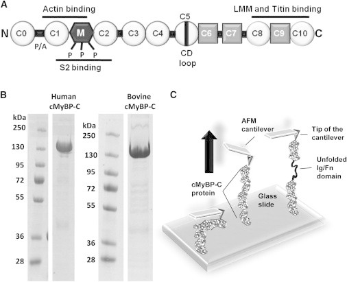
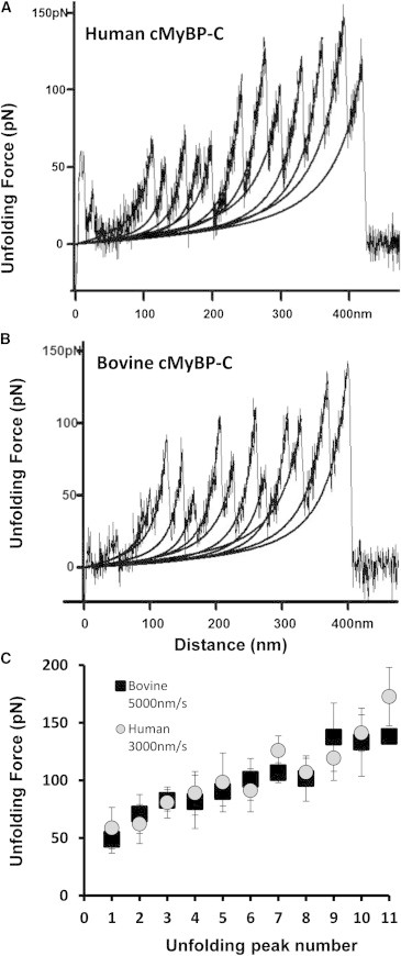
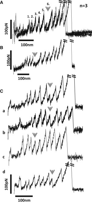
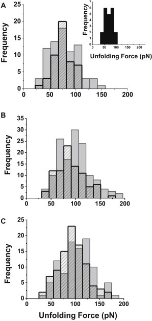
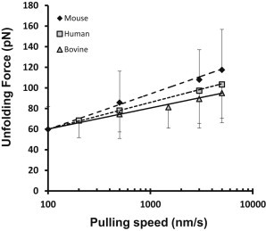
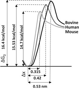
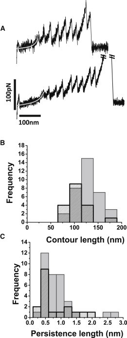
Cardiac myosin binding protein-C (cMyBP-C) is a member of the immunoglobulin (Ig) superfamily of proteins and consists of 8 Ig- and 3 fibronectin III (FNIII)-like domains along with a unique regulatory sequence referred to as the MyBP-C motif or M-domain. We previously used atomic force microscopy to investigate the mechanical properties of murine cMyBP-C expressed using a baculovirus/insect cell expression system. Here, we investigate whether the mechanical properties of cMyBP-C are conserved across species by using atomic force microscopy to manipulate recombinant human cMyBP-C and native cMyBP-C purified from bovine heart. Force versus extension data obtained in velocity-clamp experiments showed that the mechanical response of the human recombinant protein was remarkably similar to that of the bovine native cMyBP-C. Ig/Fn-like domain unfolding events occurred in a hierarchical fashion across a threefold range of forces starting at relatively low forces of ~50 pN and ending with the unfolding of the highest stability domains at ~180 pN. Force-extension traces were also frequently marked by the appearance of anomalous force drops suggestive of additional mechanical complexity such as structural coupling among domains. Both recombinant and native cMyBP-C exhibited a prominent segment ~100 nm-long that could be stretched by forces <50 pN before the unfolding of Ig- and FN-like domains. Combined with our previous observations of mouse cMyBP-C, these results establish that although the response of cMyBP-C to mechanical load displays a complex pattern, it is highly conserved across species.


Figures

Figure 1
Figure 1
Schematic diagrams showing the domain structure of cMyBP-C and an AFM experiment. (A) Domain organization of cMyBP-C showing that cMyBP-C is a multidomain protein consisting of 11 Ig- (circles) or FNIII-like domains (squares) numbered C0–C10. Sequences linking the Ig/FNIII-like domains include a sequence rich in prolines and alanines (the P/A region) located between domains C0 and C1 and the regulatory M-domain (M) located between domains C1 and C2. Phosphorylation sites (P) within the M-domain are indicated. Binding sites to myosin S2 and to actin are indicated by black lines near the N-terminus of cMyBP-C. Binding sites to light meromyosin and titin are indicated by black lines near the C-terminus. (B) Coomassie stained SDS-PAGE of recombinant human cMyBP-C and purified native bovine cMyBP-C showing protein purity. (C) Schematic diagram showing mechanical load imposed on a single cMyBP-C molecule with an AFM cantilever.
Figure 2
Figure 2
Representative force-extension curves and summary unfolding data for human and bovine cMyBP-C. Force-extension curves obtained from (A) recombinant human cMyBP-C and (B) native bovine cMyBP-C obtained at pulling speeds of 3000 and 5000 nm/s, respectively. Stretching individual cMyBP-C molecules resulted in a pattern of sawtooth peaks indicative of Ig/FNIII domain unfolding. Black lines indicate WLC model fits to the data. (C) Plots of unfolding force versus peak position (peak number) for human cMyBP-C (gray circles) and purified native bovine cMyBP-C (black squares).
Figure 3
Figure 3
Force-extension curves showing anomalous force drops. (A) Three force versus extension curves obtained from human cMyBP-C in three independent experiments are shown superimposed. Each curve shows a distinct force drop at the 6th unfolding peak (gray arrow). (B) A full-length force-extension curve of a human cMyBP-C molecule. Gray arrow indicates a force drop at the 5th unfolding peak. (C) Example force versus extension curves for human (a and b) and bovine (c and d) cMyBP-C. Gray arrows indicate force drops at various locations.
Figure 4
Figure 4
Pulling rate dependence of unfolding forces for human and bovine cMyBP-C. Unfolding-force distributions of cMyBP-C from human (gray bars) and bovine (black contours) are shown at pulling rates of (A) 500 nm/s, (B) 3000 nm/s, and (C) 5000 nm/s. Inset in A shows unfolding force of human cMyBP-C at 200 nm/s pulling rate.
Figure 5
Figure 5
Cross-species comparisons of pulling rate dependence of mean unfolding forces for cMyBP-C Ig/FNIII domains. The pulling speed dependence for the average unfolding forces of recombinant human and native bovine cMyBP-C are shown plotted at various pulling speeds and compared to recombinant mouse cMyBP-C. Data for recombinant mouse cMyBP-C were replotted from (15) to facilitate comparisons with human and bovine cMyBP-C. Solid and dashed lines are Monte Carlo simulation fits for human, bovine, and mouse cMyBP-C shown superimposed over the experimental data.
Figure 6
Figure 6
Unfolding potential diagrams from Monte Carlo simulations. Potential diagrams were drawn using activation energy (ΔG0) and unfolding potential width (Δx) parameters obtained from Monte Carlo simulations for the three different (human, bovine, and mouse) cMyBP-C proteins.
Figure 7
Figure 7
Low-compliance extensible segments of human cMyBP-C. (A) Two representative force versus extension curves for human cMyBP-C showing variable lengths of the initial extensible segments. WLC fits to the extensible segments are shown in gray. (B) Histogram of the contour length distribution of the extensible regions of human (dashed bars) (129.2 ± 25.8 nm, n = 35) and bovine cMyBP-C (gray bars) (105.1 ± 23.4 nm, n = 18). (C) Histogram of the persistence length distribution of human (dashed bars) (0.83 ± 0.51 nm, n = 35) and bovine (gray bars) (0.79 ± 0.51 nm, n = 18) cMyBP-C.

References

    1. Offer G., Moos C., Starr R. A new protein of the thick filaments of vertebrate skeletal myofibrils. Extractions, purification and characterization. J. Mol. Biol. 1973;74:653–676. - PubMed
    1. Stelzer J.E., Patel J.R., Moss R.L. Protein kinase A-mediated acceleration of the stretch activation response in murine skinned myocardium is eliminated by ablation of cMyBP-C. Circ. Res. 2006;99:884–890. - PubMed
    1. Tong C.W., Stelzer J.E., Moss R.L. Acceleration of cross-bridge kinetics by protein kinase A phosphorylation of cardiac myosin binding protein C modulates cardiac function. Circ. Res. 2008;103:974–982. - PMC - PubMed
    1. Xu Q., Dewey S., Gomes A.V. Malignant and benign mutations in familial cardiomyopathies: insights into mutations linked to complex cardiovascular phenotypes. J. Mol. Cell. Cardiol. 2010;48:899–909. - PubMed
    1. Harris S.P., Lyons R.G., Bezold K.L. In the thick of it: HCM-causing mutations in myosin binding proteins of the thick filament. Circ. Res. 2011;108:751–764. - PMC - PubMed

Publication types