

Estimated retinal ganglion cell counts in glaucomatous eyes with localized retinal nerve fiber layer defects
- PMID: 23746612
- PMCID: PMC3764310
- DOI: 10.1016/j.ajo.2013.04.015
Estimated retinal ganglion cell counts in glaucomatous eyes with localized retinal nerve fiber layer defects
Abstract
Purpose: To estimate retinal ganglion cell (RGC) losses associated with visible glaucomatous localized retinal nerve fiber layer (RNFL) defects.
Design: Observational cross-sectional study.
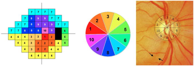
Methods: A multicenter study of 198 normal eyes (138 subjects) and 66 glaucomatous eyes (55 subjects) recruited from the Diagnostic Innovations in Glaucoma Study and the African Descent and Glaucoma Evaluation Study. All eyes underwent standard automated perimetry (SAP), spectral-domain optical coherence tomography, and fundus stereophotography within 6 months. Glaucomatous eyes were included if localized RNFL defects were detected by masked grading of stereophotographs. The number of RGCs in each sector of a structure-function map was estimated using a previously published model combining RGC estimates from SAP and spectral-domain optical coherence tomography. The estimated percentage loss of RGCs (combined structure-function index) was calculated.
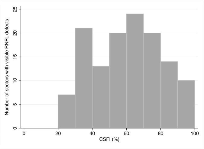
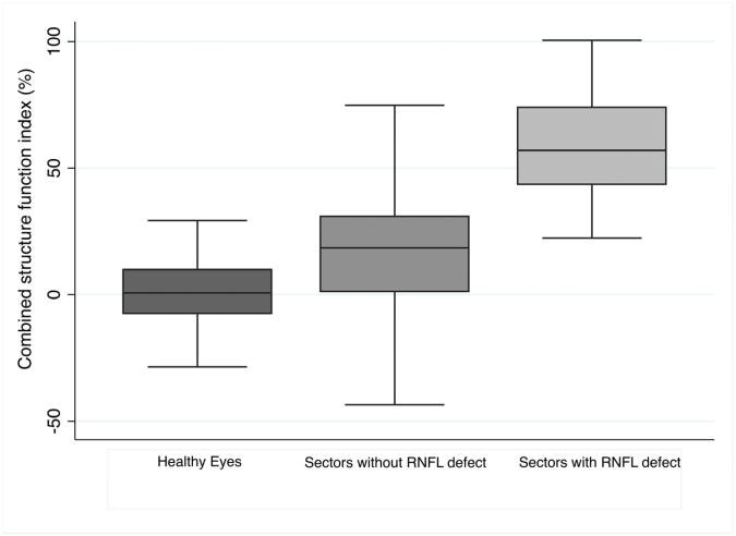
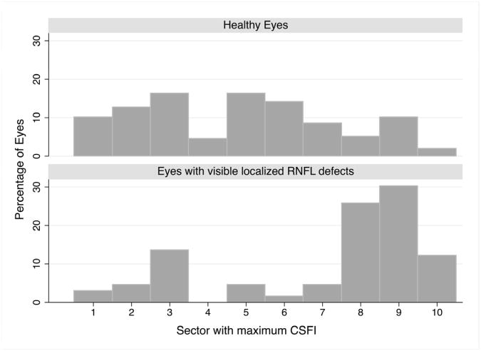
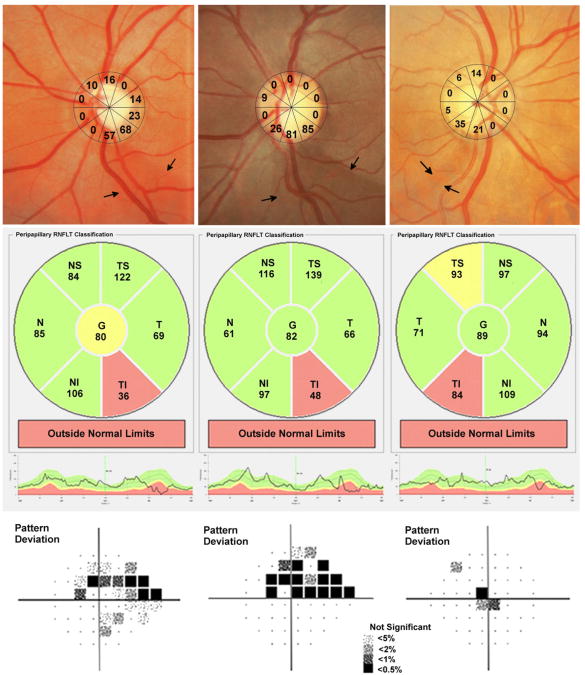
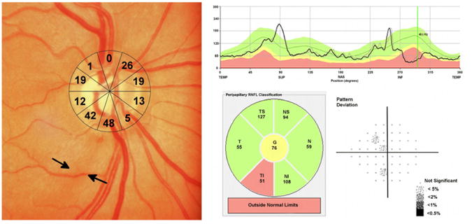
Results: In glaucomatous eyes, there were 136 sectors with visible RNFL defects and 524 sectors without visible RNFL defects. The most common sectors with visible RNFL defects were inferior and inferotemporal sectors, followed by superior and supertemporal sectors. Eyes with visible RNFL defects had a mean estimated RGC count of 657,172 cells versus 968 883 cells in healthy eyes (P < .001). The average combined structure-function index in sectors with a visible RNFL defect (59 ± 21%) was significantly higher than in sectors without a visible RNFL defect in glaucomatous eyes (15 ± 29%; P < .001) and higher than in healthy eyes (1 ± 13%; P < .001).
Conclusions: Although visible localized RNFL defects often are considered an early sign of glaucoma, this study indicates that they are likely to be associated with large neuronal losses.
Trial registration: ClinicalTrials.gov NCT00221897 NCT00221923.
Copyright © 2013 Elsevier Inc. All rights reserved.
Figures
References
-
- Quigley HA, Green WR. The histology of human glaucoma cupping and optic nerve damage: clinicopathologic correlation in 21 eyes. Ophthalmology. 1979;86(10):1803–1830. - PubMed
-
- Blumenthal EZ, Weinreb RN. Assessment of the retinal nerve fiber layer in clinical trials of glaucoma neuroprotection. Surv Ophthalmol. 2001;45(3):S305–12. discussion S332-4. - PubMed
-
- Kerrigan-Baumrind LA, Quigley HA, Pease ME, Kerrigan DF, Mitchell RS. Number of ganglion cells in glaucoma eyes compared with threshold visual field tests in the same persons. Invest Ophthalmol Vis Sci. 2000;41(3):741–748. - PubMed
-
- Quigley HA, Addicks EM. Quantitative studies of retinal nerve fiber layer defects. Arch Ophthalmol. 1982;100(5):807–814. - PubMed
Publication types
MeSH terms
Associated data
Grants and funding
LinkOut - more resources
Full Text Sources
Other Literature Sources
Medical
Miscellaneous
