. 2013 Jun 6;153(6):1366-78.
doi: 10.1016/j.cell.2013.05.015.

Regulation of organismal proteostasis by transcellular chaperone signaling

Affiliations

Regulation of organismal proteostasis by transcellular chaperone signaling

Patricija van Oosten-Hawle et al. Cell. .

Abstract

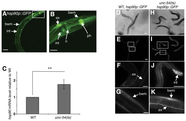
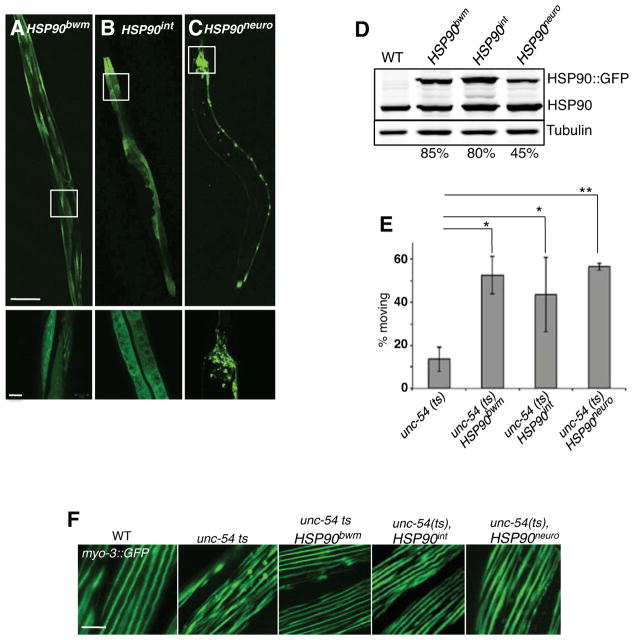
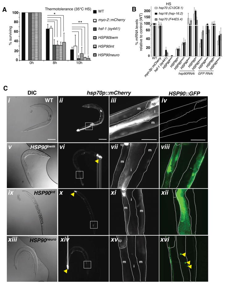
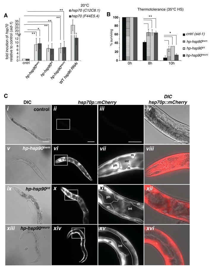
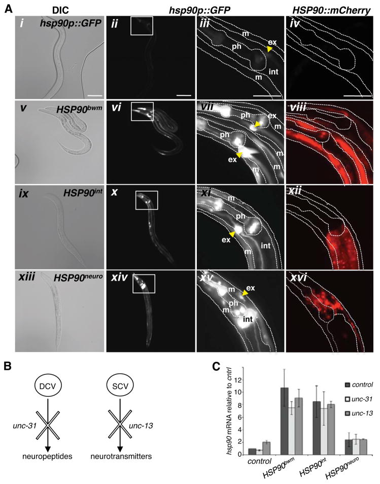
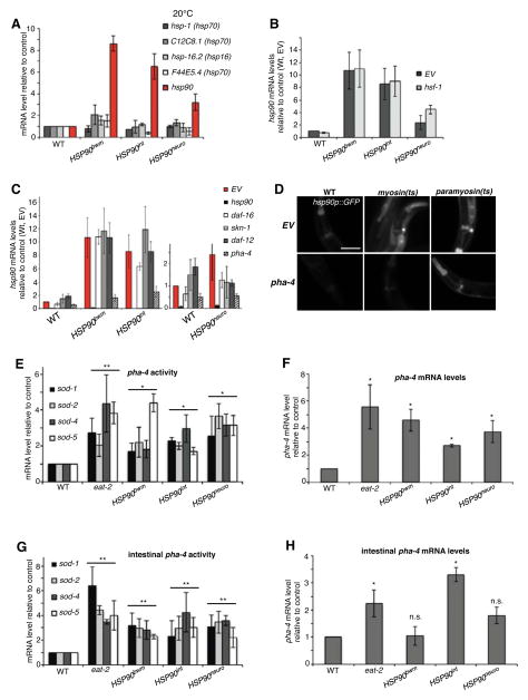
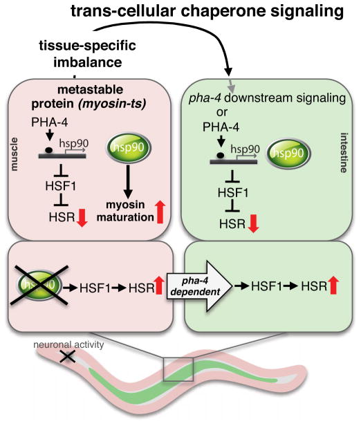
A major challenge for metazoans is to ensure that different tissues, each expressing distinctive proteomes, are nevertheless well protected at an organismal level from proteotoxic stress. We show that expression of endogenous metastable proteins in muscle cells, which rely on chaperones for proper folding, induces a systemic stress response throughout multiple tissues of C. elegans. Suppression of misfolding in muscle cells can be achieved not only by enhanced expression of HSP90 in muscle cells but as effectively by elevated expression of HSP90 in intestine or neuronal cells. This cell-nonautonomous control of HSP90 expression relies upon transcriptional feedback between somatic tissues that is regulated by the FoxA transcription factor PHA-4. This transcellular chaperone signaling response maintains organismal proteostasis when challenged by a local tissue imbalance in folding and provides the basis for organismal stress-sensing surveillance.


Figures

Figure 1
Figure 1. Tissue-specific perturbation of proteostasis is recognized across multiple tissues in a cell-non-autonomous manner
(A,B) Confocal image of a young adult animal expressing the 2.5 kb hsp90 promoter region upstream of GFP (hsp90p::GFP) at 20°C. Pronounced expression was observed in mu ltiple tissues, including pharyngeal muscle (ph), intestine (int), pharyngeal nerve ring (n), bodywall muscle (bwm) and the excretory cell (ex). Scale bar is equal to 100 μm. (B) 63x magnification of the head region. Expression is detected in the bodywall muscle (bwm), the excretory cell (ex), the pharyngeal muscle (ph), pharyngeal nerve-ring (n) and the intestine (int). Scale bar is equal to 10 μm. (C) Total mRNA levels of hsp90 in unc-54(e1301) animals relative to wild type at 15C°C. Bargraphs represent combined mean values of three independent experiments (means ± s.e.m.) **P-value < 0.01. (D–K) hsp90p::GFP reporter expression in unc-54(e1301) animals compared to wild type. Scale bar of figures (D–K) are equal to 100 μm. (F,G) 100x image of wild type expressing the hsp90 reporter in the excretory canal (ex) and bodywall muscle (bwm). (J,K) hsp90 expression in intestinal cells, bodywall muscle and excretory canal in unc-54(e1301) animals. (J) 40x image. (K) 100x image. All fluorescent images were taken at equal exposure times. See also Figure S1.
Figure 2
Figure 2. Tissue-specific increased levels of HSP90 improves the organismal folding environment of myosin(ts) mutants
(A–C) Confocal images of young adult C. elegans animals overexpressing HSP90::GFP in the (A) bodywall muscle (HSP90bwm), (B) the intestine (HSP90int) or (C) the neurons (HSP90neuro) (scale bar = 100 μm), with 63x magnifications of selected regions (Scale bar = 10 μm). (D) Western Blot analysis of young adult animals overexpressing HSP90 using an anti-C.elegans HSP90 antibody. Levels of HSP90::GFP are normalized to the loading control (tubulin) and relative to endogenous HSP90. (E) Percentage of young adult animals expressing ts myosin [unc-54 (e1301)] alone or in the presence of bodywall muscle-specific HSP90::GFP overexpression (unc-54(ts),HSP90bwm), intestinal overexpression (unc-54(ts),HSP90int), or neuronal overexpression (unc-54(ts),HSP90neuro) showing movement after exposure to restrictive temperature (25°C) for 12 – 24 hours. n = 20 adult animals per strain per experiment. Bargraphs represent the combined results of 3 independent experiments. Error bars = ±SEM. *P-value < 0.05. **P-value < 0.01. (F) Confocal images of the bodywall muscle of age-synchronized wild type, unc-54(e1301), unc-54(e1301),HSP90bwm, unc-54(e1301),HSP90int, and unc-54(ts),HSP90neuro animals after exposure to restrictive temperature (25ºC) for 12 hours, using myo-3::GFP for visualization. Scalebar = 20 μm. See also Figure S2.
Figure 3
Figure 3. Elevated tissue-specific HSP90 levels repress the HSR at an organismal level
(A) Thermosensitivity of young adult animals (n = 100) with indicated genotypes exposed to 35°C heat stress. *P-value < 0.05. **P-value < 0.01. (B) Total mRNA levels of hsp70 (C12C8.1 and F44E5.4) and hsp16 (hsp-16.2) after heat shock (1h at 33°C) in young adult myo-2p::mCherry, hsf-1 (sy441) mutant, HSP90neuro, HSP90int, HSP90bwm animals and upon RNAi-mediated GFP or hsp90 knockdown prior to heat shock in the transgenic HSP90 lines, relative to wild type. (A and B) Bargraphs represent combined mean values of three independent experiments (means ± s.e.m.) (C) DIC Nomarski images of (i) wild type, (v) HSP90bwm, (ix) HSP90int and (xiii) HSP90neuro animals expressing the hsp70p::mCherry reporter. Expression of the hsp70p::mCherry reporter 7 hours after heat shock (33°C, 1h) in representative (ii) wild type and in (vi) HSP90bwm, (x) HSP90int and (xiv) HSP90neuro animals. Yellow arrow in vi, x, and xiv indicates the pharyngeal myo-2::mCherry co-injectionmarker, present in all transgenic HSP90::GFP lines. (iii,vii,xi,xv) 20x magnifications of the posterior region of (iii) wild type and HSP90 overexpression lines (vii, xi, xv), indicating hsp70 induction in spermatheca (sp), bodywall muscle (m) and the intestine (i). (iv, viii, xii, xvi) HSP90::GFP expression in true color. Yellow arrows in (xvi) indicate neuronal cells expressing HSP90::GFP. (i–xvi) Scale = 100 μm. See also Figure S3.
Figure 4
Figure 4. Tissue-specific knockdown of hsp90 cell-non-autonomously induces the HSR
(A) Bodywall-muscle, intestine- and neuron-specific hsp90 RNAi induces basal levels of hsp70 (C12C8.1 and F44E5.4) expression at 20°C, compared to control animals (sid-1). Wild type animals allow import of dsRNA from surrounding tissues, leading to higher induction of organismal hsp70 than in the tissue-specific knockdown lines. Bargraphs represent combined mean values of three independent experiments (means ± s.e.m.) **P-value < 0.05. (B) Thermosensitivity of young adult animals (n = 100) expressing the indicated tissue-specific hp-hsp90 construct exposed to 35 °C heat stress. Bargraphs represent combined mean values of three independent experiments (means ± s.e.m.) **P-value < 0.01. *P-value < 0.05. (C) Tissue-specific knockdown of hsp90 induces expression of the hsp70 reporter (hsp70p::mCherry) at 20°C. DIC images of synchronized young adult (i) control animals (sid-1), (v) hp-hsp90bwm, (ix) hp-hsp90int, and (xiii) hp-hsp90neuro animals expressing the hsp70 reporter. Expression of the hsp70p::mCherry in (ii) sid-1 control animals, (vi) hp-hsp90bwm, (x) hp-hsp90int, and (xiv) hp-hsp90neuro. (iii, vii, xi, xv) 20x magnification of control (iii) and tissue-specific hsp90 knockdown lines (vii, xi, xv) indicating expression of hsp70p::mCherry in the pharynx (ph), intestine (int) and bodywall muscle (m). (iv, viii, xii, xvi) Overlay of DIC Nomarski and hsp70p::mCherry (red). Scalebars = 100 μm. See also Figure S4.
Figure 5
Figure 5. Coordination of cell-non-autonomous hsp90 expression is regulated independent of neural control
(A) Tissue-selective increased levels of HSP90 up-regulates expression of the transcriptional hsp90p::GFP reporter at normal conditions (20°C). DIC images o f (i) wildtype, (v) HSP90bwm, (ix) HSP90int and (xiii) HSP90neuro expressing the hsp90p::GFP reporter. hsp90 reporter expression in representative whole animals (ii, vi, x, xiv) and magnified head region (iii, vii, xi, xv) in (ii, iii) wild type, (vi, vii) HSP90bwm, (x, xi) HSP90int, and (xiv, xv) HSP90neuro, indicating hsp90p::GFP in the pharynx (ph), the intestine (int), bodywall muscle (m) and excretory cell (ex). (iv, viii, xii, xvi) HSP90::mCherry (red) expression in true color. Yellow arrows in (iii, vii, xi and xv) indicate hsp90p::GFP expression in the excretory cell. Scalebars = 100 μm.(B) Schematic representation of the major modes of neuro-secretion in C. elegans, regulated via either dense core vesicles (DCV) through unc-31 or via small core vesicles (SCV) through unc-13. (C) Total hsp90 mRNA levels of HSP90 overexpression lines in an unc-31 deletion mutant background (white bars) or crossed to an unc-13 deletion mutant (light grey bars) relative to control animals (dark grey bars). Bargraphs represent combined mean values of three independent experiments (means ± s.e.m.). See also Figure S5.
Figure 6
Figure 6. A PHA-4 – dependent transcriptional feedback coordinates cell-non-autonomous hsp90 expression
(A) Total mRNA levels of hsp-1 (constitutive hsp70), hs-inducible hsp70 (C12C8.1 and F44E5.4), small heat shock protein hsp16 (hsp-16.2) and hsp90 in HSP90 overexpression lines at 20°C relative to wild type. The slightly increased hsp levels in the HSP90bwm may be indicative of the higher sensitivity of muscle cells to proteostatic perturbation, in line with the observations on tissue-specific hsp90 knockdown, where the HSR is primarily induced in the bodywall muscle. (B) Organismal hsp90 expression in HSP90 overexpression lines is independent of hsf-1. hsp90 mRNA levels in control (EV) and animals fed with hsf-1 RNAi. Whereas hsp90 expression in wild type is hsf-1 dependent, RNAi-mediated knockdown of hsf-1 leaves organismal hsp90 levels in HSP90bwm, HSP90int or HSP90neuro unchanged. (C) pha-4 RNAi decreases elevated hsp90 expression in the overexpression lines. (D) hsp90p::GFP reporter expression in myosin (ts, e1157) or paramyosin (ts, e1402) mutants is reduced during pha-4 RNAi when compared to control RNAi (EV). Scalebar = 50 μm. (E) pha-4 activity is increased in the HSP90 overexpression lines. mRNA levels of pha-4 regulated genes sod-1, sod-2, sod-4 and sod-5 are induced in eat-2(ad1113) mutants, HSP90bwm, HSP90int and HSP90neuro animals relative to wild type. *P-value < 0.05. **P-value < 0.02. (F) pha-4 mRNA expression levels in eat-2 and HSP90 overexpression lines are upreguated, relative to the wild type control. *P-value < 0.05. (G) pha-4 activity in intestinal cells of eat-2(ad1113), HSP90bwm, HSP90int and HSP90neuro animals relative to wild type. **P-value < 0.02. (H) pha-4 mRNA expression levels are induced in the intestines of eat-2 mutants and HSP90int (=signaling tissue), but not in the intestines of HSP90bwm or HSP90neuro (=recipient tissue). *P-value < 0.05. (n.s. = not significant). (A – H) All bar graphs represent combined mean values of three independent experiments (3 biological replicates) (means ± s.e.m.). See also Figure S6.
Figure 7
Figure 7. Model for the cell non-autonomous regulation of HSP90 expression in C. elegans.
An imbalance of the proteostasis network through the presence of metastable myosin increases the expression of HSP90 in muscle cells but also in different cell-types, such as the intestine. This is regulated by PHA-4 and communicated through trans-cellular chaperone signaling to other tissues, independent of neural activity. Increased pha-4 activity is required in the signaling and receiving tissue, however pha-4 may act from a distance to regulate gene expression in the receiving tissue via a down-stream signaling cascade. The resulting highly abundant HSP90 levels in the entire animal are beneficial for myosin folding in unc-54(ts) mutants, but can become detrimental during severe heat shock, due to cell non-autonomous repression of HSF-1 transcriptional activity. Likewise, a tissue-specific perturbation through reduced hsp90 levels (lower panel) leads to induction of the HSR in the same and recipient tissues. Transduction of the response to the recipient tissue is also pha-4 dependent.

Comment in

References

    1. Akerfelt M, Morimoto RI, Sistonen L. Heat shock factors: integrators of cell stress, development and lifespan. Nat Rev Mol Cell Biol. 2010;11:545–555. - PMC - PubMed
    1. Altun-Gultekin ZaHDH. Coelomocyte System. WormAtlas 2009
    1. Barral JM, Hutagalung AH, Brinker A, Hartl FU, Epstein HF. Role of the myosin assembly protein UNC-45 as a molecular chaperone for myosin. Science. 2002;295:669–671. - PubMed
    1. Belting M, Wittrup A. Nanotubes, exosomes, and nucleic acid-binding peptides provide novel mechanisms of intercellular communication in eukaryotic cells: implications in health and disease. The Journal of Cell Biology. 2008;183:1187–1191. - PMC - PubMed
    1. Ben-Zvi A, Miller EA, Morimoto RI. Collapse of proteostasis represents an early molecular event in Caenorhabditis elegans aging. Proc National Acad Sci U S A. 2009;106:14914–14919. - PMC - PubMed

Publication types

MeSH terms