. 2013 Nov;9(8):1235-44.
doi: 10.1016/j.nano.2013.05.010. Epub 2013 Jun 6.

A comprehensive analysis of transfection-assisted delivery of iron oxide nanoparticles to dendritic cells

Affiliations

A comprehensive analysis of transfection-assisted delivery of iron oxide nanoparticles to dendritic cells

Shinji Toki et al. Nanomedicine. 2013 Nov.

Abstract

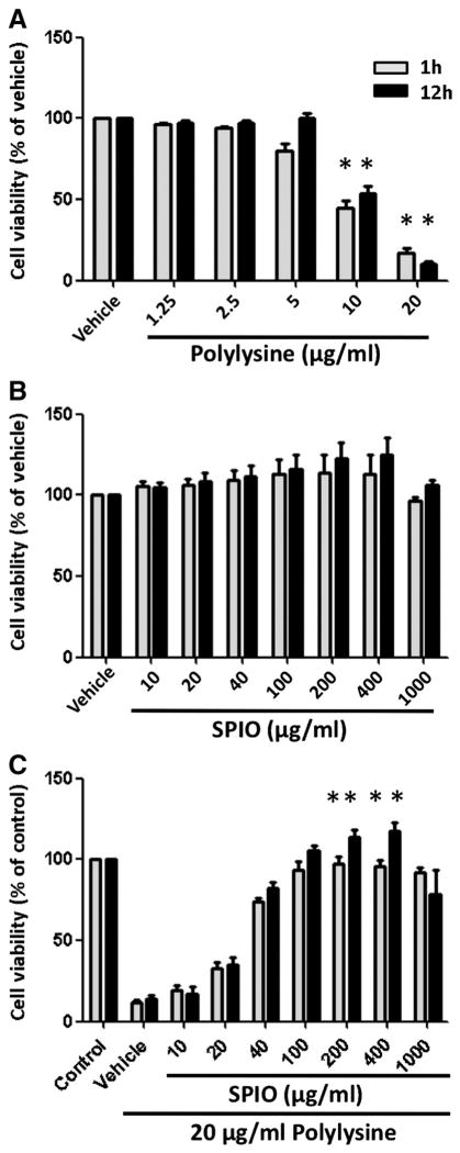
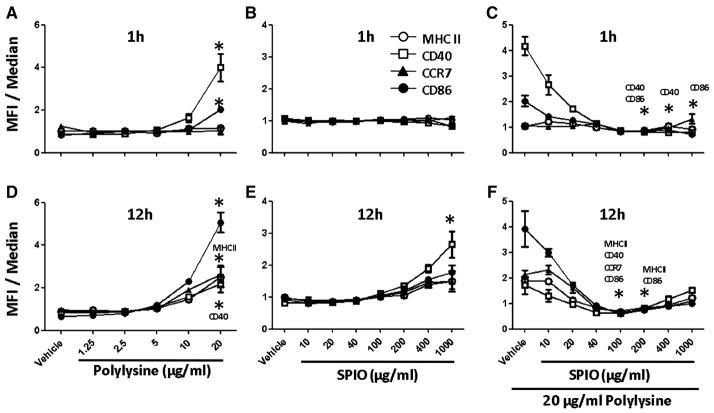
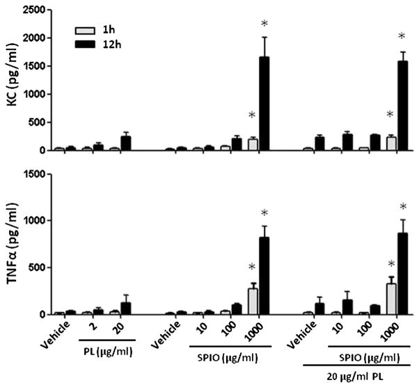
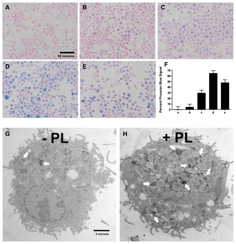
Polylysine (PL) has been used to facilitate dendritic cell (DC) uptake of super paramagnetic iron oxide (SPIO) nanoparticles for use in magnetic resonance imaging (MRI). In this work, we examined the effect of PL on cell toxicity and induction of cell maturation as manifested by the up-regulation of surface molecules. We found that PL became toxic to bone marrow-derived DCs (BMDCs) at the 10 μg/ml threshold. Incubation of BMDCs with 20 μg/ml of PL for 1h resulted in approximately 90% cell death. However, addition of SPIO nanoparticles rescued DCs from PL-induced death as the combination of SPIO with PL did not cause cytotoxicity until the PL concentration was 1000 μg/ml. Prolonged exposure to PL induced BMDC maturation as noted by the expression of surface molecules such as MHC class II, CD40, CCR7 and CD86. However, the combination of SPIO and PL did not induce BMDC maturation at 1h. However prolonged exposure to SPIO nanoparticles induced CD40 expression and protein expression of TNFα and KC. The data suggest that the use of PL to enhance the labeling of DCs with SPIO nanoparticles is a dedicated work. Appropriate calibration of the incubation time and concentrations of PL and SPIO nanoparticles is crucial to the development of MRI technology for noninvasive imaging of DCs in vivo.

From the clinical editor: The authors of this study present detailed data on toxicity and efficiency of polylysine-facilitated uptake of USPIO-s by dendritic cells for cell-specific MR imaging.

Keywords: Dendritic cells; Iron oxide nanoparticles; Nanotechnology; Polylysine; Transfection.


Figures

Figure 1
Figure 1
Viability of BMDCs incubated with PL, SPIO nanoparticle or both. Immature, BMDCs were grown in the absence (vehicle) or presence of increasing concentrations of (A) PL, (B) SPIO nanoparticles, and (C) both PL and SPIO nanoparticles for periods of 1 and 12 h. Control means BMDCs alone. Data were normalized to the vehicle or control. Columns, mean from four experiments; bars, SE. *, Statistical significance (P < 0.05), compared with the vehicle or control.
Figure 2
Figure 2
Cell surface phenotype as determined by flow cytometric analysis of unlabeled BMDCs (vehicle), PL-labeled (A, D), SPIO-labeled cells (B, E) or PL-labeled cells in the presence of increasing concentrations of SPIO nanoparticles (C, F). Following 1 and 12 h of incubation with PL, SPIO nanoparticles or both, 1.0 × 10⁶ cells were stained with monoclonal antibodies for antigen presenting receptors MHC-II as well as costimulatory molecules such as CD40, CD86 and chemokine receptor CCR7. Data from the histograms were normalized to the median and compiled into the charts (n = 4 per time point per group). *, Statistical significance P < 0.05, compared to the vehicle.
Figure 3
Figure 3
Viability and cell surface phenotype of BMDCs incubated with dextran T-10 or a combination of PL and dextran T-10. Immature BMDCs were grown in the absence (vehicle) or presence of increasing concentrations of dextran T-10 (A, B) or a combination of PL and dextran T-10 for 12 h (C, D). The control was comprised of BMDCs alone. Data that appear in the bar graph were normalized to the vehicle or control. Data that appear in the line graph were normalized to the median. Each value represents the mean from four experiments. Bars represent SE.
Figure 4
Figure 4
Analysis of the cytokine profiles in the cell culture medium 1 and 12 h after treating BMDCs with PL, SPIO nanoparticles or both. Each value represents the mean from four experiments. Bars represent SE. *, Statistical significance P < 0.05, compared to the vehicle.
Figure 5
Figure 5
Prussian blue staining was used to assess the effect of PL-induced internalization of SPIO nanoparticles by immature BMDCs that had been labeled for 12 h (nuclear fast red counterstain; magnification at 600×). Non-labeled BMDCs (A), 500 μg/ml SPIO (B), 500 μg/ml SPIO + 10 μg/ml PL mixture (C), 500 μg/ml SPIO nanoparticles coated with PL during a 3-h preincubation (D), 100 μg/ml SPIO + 20 μg/ml PL mixture (E). Quantitative analysis of the uptake of SPIO nanoparticles shown in A, B, C, D and E by MATLAB (F). PL-mediated delivery of SPIO nanoparticles was confirmed by TEM. Scale bars 2 μm (G and H); * Statistical significance P < 0.05.

References

    1. Pham W, Kobukai S, Hotta C, Gore JC. Dendritic cells: therapy and imaging. Expert Opin Biol Ther. 2009;9:539–64. - PubMed
    1. Grolleau A, Sloan A, Mule JJ. Dendritic cell-based vaccines for cancer therapy. In: Samir Khleif., editor. Tumor immunology and cancer vaccines. 2005. pp. 181–205. - PubMed
    1. Mohty M, Olive D, Gaugler B. Leukemic dendritic cells: potential for therapy and insights towards immune escape by leukemic blasts. Leukemia. 2002;16:2197–204. - PubMed
    1. Kobukai S, Baheza R, Cobb JG, Virostko J, Xie J, Gillman A, et al. Magnetic nanoparticles for imaging dendritic cells. Magn Reson Med. 2010;63:1383–90. - PMC - PubMed
    1. Kupiec-Weglinski JW, Austyn JM, Morris PJ. Migration patterns of dendritic cells in the mouse. Traffic from the blood, and T cell-dependent and -independent entry to lymphoid tissues. J Exp Med. 1988;167:632–45. - PMC - PubMed

Publication types

MeSH terms