. 2013 Aug;183(2):450-8.
doi: 10.1016/j.ajpath.2013.04.031. Epub 2013 Jun 7.

Compartmentalization of immune responses during Staphylococcus aureus cranial bone flap infection

Affiliations

Compartmentalization of immune responses during Staphylococcus aureus cranial bone flap infection

Joseph Cheatle et al. Am J Pathol. 2013 Aug.

Abstract

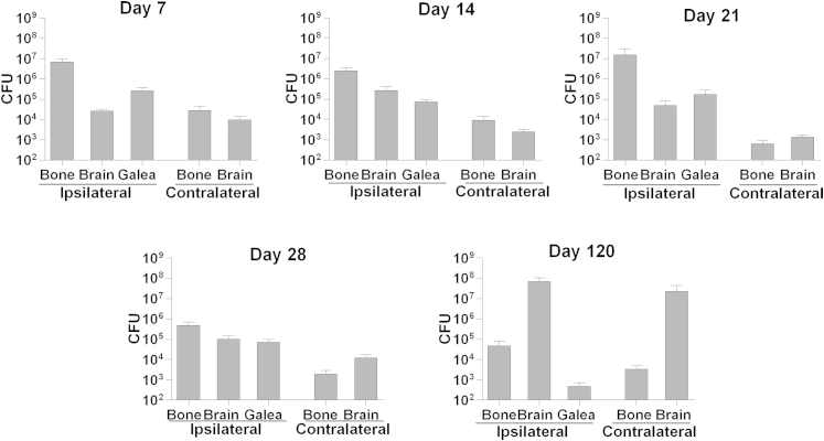
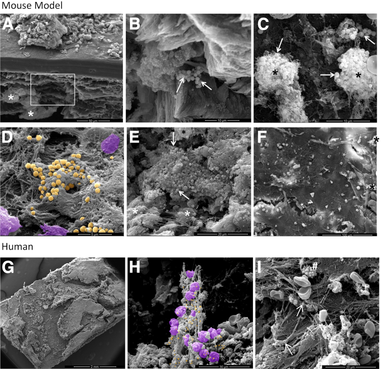
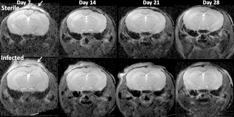
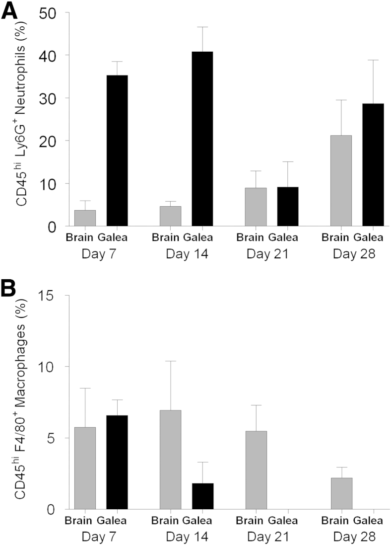
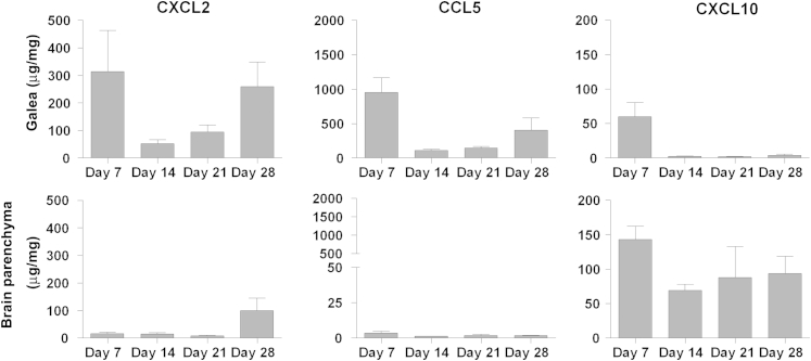
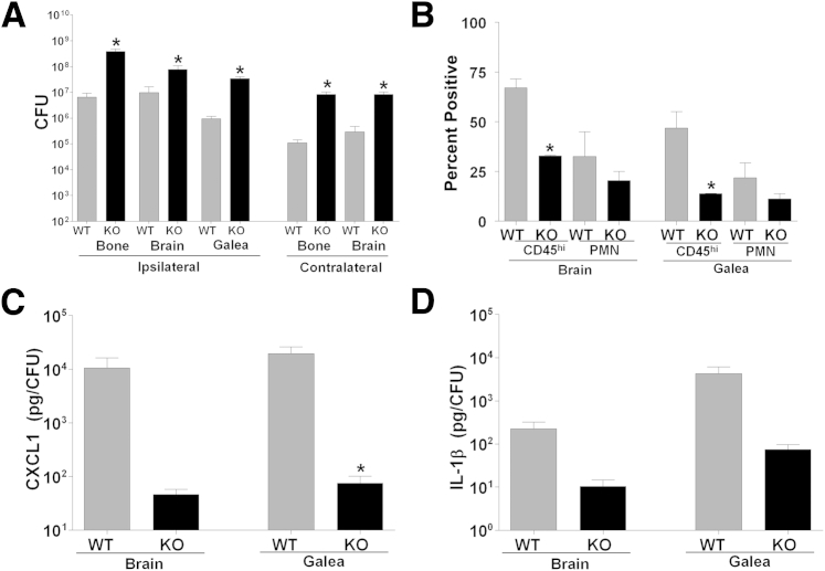
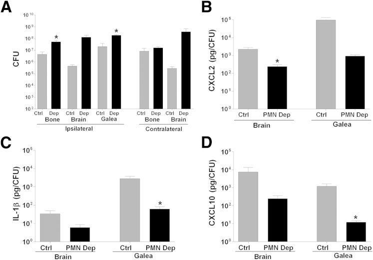
Decompressive craniectomy is often required after head trauma, stroke, or cranial bleeding to control subsequent brain swelling and prevent death. The infection rate after cranial bone flap replacement ranges from 0.8% to 15%, with an alarming frequency caused by methicillin-resistant Staphylococcus aureus, which is problematic because of recalcitrance to antibiotic therapy. Herein we report the establishment of a novel mouse model of S. aureus cranial bone flap infection that mimics several aspects of human disease. Bacteria colonized bone flaps for up to 4 months after infection, as revealed by scanning electron microscopy and quantitative culture, demonstrating the chronicity of the model. Analysis of a human cranial bone flap with confirmed S. aureus infection by scanning electron microscopy revealed similar structural attributes as the mouse model, demonstrating that it closely parallels structural facets of human disease. Inflammatory indices were most pronounced within the subcutaneous galeal compartment compared with the underlying brain parenchyma. Specifically, neutrophil influx and chemokine expression (CXCL2 and CCL5) were markedly elevated in the galea, which demonstrated substantial edema on magnetic resonance images, whereas the underlying brain parenchyma exhibited minimal involvement. Evaluation of immune mechanisms required for bacterial containment and inflammation revealed critical roles for MyD88-dependent signaling and neutrophils. This novel mouse model of cranial bone flap infection can be used to identify key immunologic and therapeutic mechanisms relevant to persistent bone flap infection in humans.


Figures

Figure 1
Figure 1
S. aureus colonization of cranial bone flaps leads to persistent infection in multiple compartments. Infected bone flaps (105 CFU) were reinserted after craniectomy. Mice were sacrificed at the indicated times after infection, and the numbers of viable bacteria on the ipsilateral bone flap, galea, and adjacent brain parenchyma were determined. Equivalent regions were also sampled from the contralateral hemisphere. Animals that received sterile bone flaps tested consistently negative for bacterial growth, and their data are not shown. Results are representative of combined data from 13 to 15 mice over the course of three independent experiments.
Figure 2
Figure 2
S. aureus forms complex structures during cranial bone flap infection. SEM analysis of infected (A–E) and sterile (F) bone flaps from mice at day 7 after reinsertion into the cranium. Inset in A depicts the region magnified in B, highlighting a focus of S. aureus associated with the ventral bone flap surface. C: Cocoon-like structures harboring numerous bacteria. D and E: Various complex tertiary formations that were frequently observed on the bone flap surface. G–I: SEM images of a bone flap from a patient with confirmed S. aureus infection revealed similarities to the mouse model. G: Overview of the human bone flap surface reveals an irregular pattern of material deposition. H: Tertiary structure formed by interactions between leukocytes and S. aureus. I: Fiber deposition on the human bone flap surface shows remarkable similarity to the mouse model (D). Images in D and H have been pseudo-colored to highlight bacteria (gold) and leukocytes (purple). Bacteria are indicated by arrows, and host cells are depicted by either black or white asterisks. Hash mark in I indicates a crenated red blood cell artifact, which is also apparent in H. Scale bars: 50 μm (A); 10 μm (B and C); 5 μm (D); 20 μm (E and I); 100 μm (F); 2 mm (G); and 30 μm (H).
Figure 3
Figure 3
MRI analysis reveals prominent galeal inflammation during S. aureus cranial bone flap infection. Sterile or infected (105 CFU) bone flaps were reinserted after craniectomy, and mice were subjected to MRI analysis at weekly intervals to day 28 after flap reinsertion into the cranium. Arrows indicate the region of bone flap replacement in T2-weighted RARE images from the same mouse over time. Results are representative of eight individual mice per group.
Figure 4
Figure 4
Differential recruitment of neutrophils and macrophages into distinct compartments during cranial bone flap infection. Infected bone flaps (105 CFU) were reinserted after craniectomy. Mice were sacrificed at weekly intervals extending to day 28 after infection, and percentages of neutrophils (A) and macrophages (B) infiltrating the ipsilateral galea and adjacent brain parenchyma were determined by FACS. Results were combined from three independent experiments.
Figure 5
Figure 5
Chemokines are preferentially expressed in distinct compartments during S. aureus cranial bone flap infection. Infected bone flaps (105 CFU) were reinserted after craniectomy. Mice were sacrificed at weekly intervals extending to day 28 after infection, and CXCL2, CCL5, and CXCL10 expression in the ipsilateral galea and adjacent brain parenchyma was determined using multianalyte bead arrays. Results are normalized to the amount of total protein to correct for differences in tissue sampling size.
Figure 6
Figure 6
MyD88-dependent signaling is important for bacterial containment and proinflammatory mediator secretion during S. aureus cranial bone flap infection. Infected bone flaps (105 CFU) were reinserted in MyD88 KO and WT mice after craniectomy. Animals were sacrificed at day 2 after infection, and bacterial burdens (A), CD45+ leukocyte and neutrophil (PMN) infiltrates (B), and CXCL1 (C) and IL-1β (D) expression associated with the ipsilateral galea and adjacent brain parenchyma were evaluated. CXCL1 and IL-1β levels were determined using multianalyte bead arrays and are normalized to bacterial burdens. Results are representative of two independent experiments. P < 0.05.
Figure 7
Figure 7
Neutrophils are critical for controlling bacterial burdens and eliciting proinflammatory mediator expression during cranial bone flap infection. Infected bone flaps (105 CFU) were reinserted in neutrophil-depleted (PMN Dep) and control (Ctrl) mice after craniectomy. Animals were sacrificed at day 2 after infection, and bacterial burdens (A) and CXCL2 (B), IL-1β (C), and CXCL10 (D) expression associated with the ipsilateral galea and adjacent brain parenchyma were evaluated. CXCL2, IL-1β, and CXCL10 levels were determined using multi-analyte bead arrays and are normalized to bacterial burdens. Results are representative of two independent experiments. P < 0.05.

References

    1. Bhaskar I.P., Zaw N.N., Zheng M., Lee G.Y. Bone flap storage following craniectomy: a survey of practices in major Australian neurosurgical centres. ANZ J Surg. 2011;81:137–141. - PubMed
    1. Dashti S.R., Baharvahdat H., Spetzler R.F., Sauvageau E., Chang S.W., Stiefel M.F., Park M.S., Bambakidis N.C. Operative intracranial infection following craniotomy. Neurosurg Focus. 2008;24:E10. - PubMed
    1. Baumeister S., Peek A., Friedman A., Levin L.S., Marcus J.R. Management of postneurosurgical bone flap loss caused by infection. Plast Reconstr Surg. 2008;122:195e–208e. - PubMed
    1. Hammon W.M., Kempe L.G. Methyl methacrylate cranioplasty: 13 years experience with 417 patients. Acta Neurochir (Wien) 1971;25:69–77. - PubMed
    1. Honeybul S., Ho K.M. Long-term complications of decompressive craniectomy for head injury. J Neurotrauma. 2011;28:929–935. - PubMed

Publication types

MeSH terms