. 2013 Sep;749(1-2):49-57.
doi: 10.1016/j.mrfmmm.2013.05.004. Epub 2013 Jun 6.

The rate of spontaneous mutations in human myeloid cells

Affiliations

The rate of spontaneous mutations in human myeloid cells

David J Araten et al. Mutat Res. 2013 Sep.

Abstract

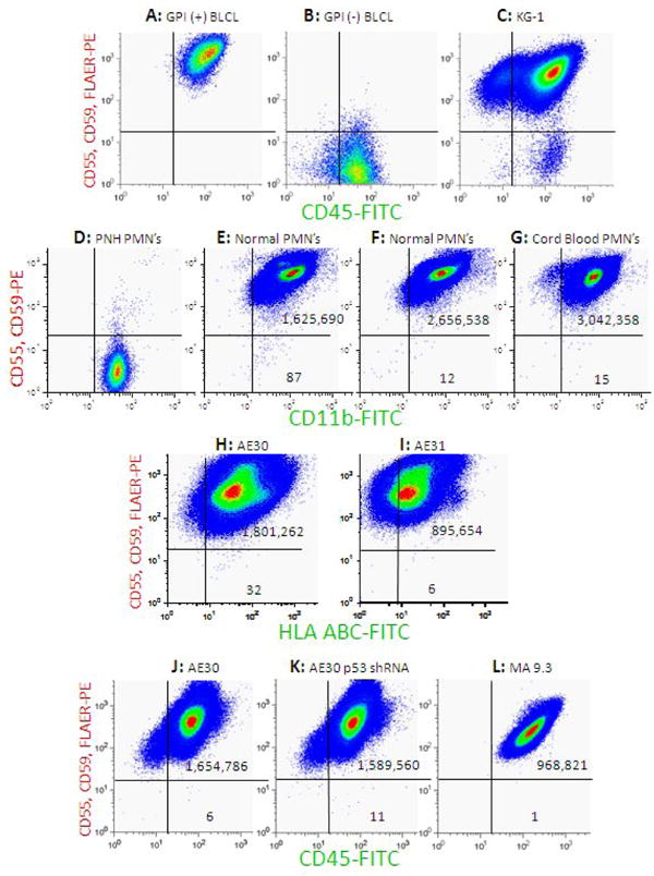
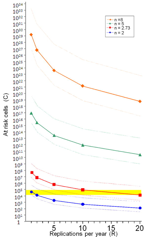
The mutation rate (μ) is likely to be a key parameter in leukemogenesis, but historically, it has been difficult to measure in humans. The PIG-A gene has some advantages for the detection of spontaneous mutations because it is X-linked, and therefore only one mutation is required to disrupt its function. Furthermore, the PIG-A-null phenotype is readily detected by flow cytometry. Using PIG-A, we have now provided the first in vitro measurement of μ in myeloid cells, using cultures of CD34+ cells that are transduced with either the AML-ETO or the MLL-AF9 fusion genes and expanded with cytokines. For the AML-ETO cultures, the median μ value was ∼9.4×10(-7) (range ∼3.6-23×10(-7)) per cell division. In contrast, few spontaneous mutations were observed in the MLL-AF9 cultures. Knockdown of p53 or introduction of mutant NRAS or FLT3 alleles did not have much of an effect on μ. Based on these data, we provide a model to predict whether hypermutability must occur in the process of leukemogenesis.

Keywords: AML; B-lymphoblastoid cell lines; BLCLs; GPI; GPI-linked proteins; Human myeloid cultures; Mutation rate; Myeloid leukemia.; PIG-A gene; PNH; Spontaneous somatic mutations; acute myelogenous leukemia; glycosylphosphatidylinositol; paroxysmal nocturnal hemoglobinuria.


Conflict of interest statement

Conflicts of interest: The authors have no relevant conflicts of interest to disclose

Figures

Figure 1
Figure 1
Bivariate pseudocolor dot plot analysis demonstrating GPI (+) and GPI (-) cells. (A) BLCL from a normal donor. These cells express GPI-anchored surface proteins such as CD55 and CD59 and bind to the FLAER reagent and also express transmembrane proteins such as CD45; (B) Control GPI (-) BLCL cells from a patient with PNH do not express CD55 or CD59 and do not take up the FLAER reagent but do express transmembrane proteins; (C) KG-1, an AML-derived cell line. There is a large subpopulation of spontaneously arising cells with the GPI (-) phenotype (lower right quadrant); (D) Granulocytes from a patient with PNH, which do not express CD55 or CD59 but do express transmembrane proteins such as CD11b; (E and F) Granulocytes from two normal donors. The vast majority of the cells are GPI (+) and appear in the upper right quadrant, and a small population of spontaneously arising GPI (-) cells are seen in the lower right quadrant. The mutant frequency is 54 × 10-6 and 4.5 × 10⁻⁶ respectively, representing the high and low end of our previously reported range in normal donors. (G) Granulocytes from an umbilical cord blood sample. As in the adult samples, the vast majority of the cells are GPI (+) and appear in the upper right quadrant, and a small population of spontaneously arising GPI (-) cells are seen in the lower right quadrant at a frequency of 4.9 × 10⁻⁶. (H and I) AE30 and AE31-derived myeloid cell cultures. As for mature granulocytes, the vast majority are GPI (+) and there is a small population of spontaneously arising GPI (-) cells at a frequency of 17.8 × 10⁻⁶ and 6.7 × 10⁻⁶ respectively (J) AE30-derived myeloid cultures, shown after expansion after sorting to eliminate pre-existing GPI (-) cells. New GPI (-) cells have spontaneously arisen in culture and appear in the lower right quadrant (see Table 1 for calculations of μ); (K) AE30-derived myeloid cells with an shRNA targeting p53, analyzed after expansion after sorting. f and μ are only modestly higher in this example than for the AE30-derived cell line with normal p53, shown in panel J. (L) CD34+ cell line transduced with the MLL-AF9 fusion gene, analyzed after expansion after sorting. This cell line has very low f and μ values.
Figure 2
Figure 2
The number of stem or progenitor cells (C) that must be capable of giving rise to AML (shown on a log scale) is calculated for different estimates of their annual rate of cell replication (R) based on different estimates of n. The solid lines represent the calculations for C and R based on the mean μ value for the AE cell lines (∼12 × 10-7 per cell division), and the dotted lines represent the C and R values based on the lower and upper range of the calculated μ values for the AE cell lines (3.6 × 10⁻⁷ and 23 × 10-7). The yellow highlighted region indicates the estimated number of hematopoietic stem cells (14,000 to 80,000 [1,2]). If n = 8, C is greater than the number of cells in the body, indicating that hypermutability must occur at some point in the process of malignant transformation. Only in the case where n = 2 is it possible that AML could arise from within a true hematopoietic stem cell without any elevation in μ.

Similar articles

Cited by

References

    1. DePinho R. The age of cancer. Nature. 2000;408:248–254. - PubMed
    1. Edwards B, Howe H, Ries L, Thun M, Rosenberg H, Yancik R, Wingo P, Jemal A, Feigal E. Annual Report to the Nation on the Status of Cancer 1973-1999, Featuring Implications of Age and Aging on US Cancer Burden. Cancer. 2002;94:2766–2792. - PubMed
    1. Moorman A, Roman E, Cartwright R, Morgan G. Age-specific incidence rates for cytogenetically-defined subtypes of acute myeloid leukaemia. British Journal of Cancer. 2002;86:1061–1063. - PMC - PubMed
    1. Albertini R, Nicklas J, O'Neill J, Robison S. In vivo somatic mutations in humans: measurement and analysis. Annu Rev Genet. 1990;24:305–326. - PubMed
    1. Albertini R, Castle K, Borcherding W. T cell cloning to detect the mutant 6-thioguanine-resistant lymphocytes present in human peripheral blood. PNAS. 1982;79:6617–6621. - PMC - PubMed

Publication types

MeSH terms

Substances