Review
. 2014 Feb;1840(2):847-75.
doi: 10.1016/j.bbagen.2013.05.040. Epub 2013 Jun 6.

Sulfenic acid chemistry, detection and cellular lifetime

Affiliations
Review

Sulfenic acid chemistry, detection and cellular lifetime

Vinayak Gupta et al. Biochim Biophys Acta. 2014 Feb.

Abstract

Background: Reactive oxygen species-mediated cysteine sulfenic acid modification has emerged as an important regulatory mechanism in cell signaling. The stability of sulfenic acid in proteins is dictated by the local microenvironment and ability of antioxidants to reduce this modification. Several techniques for detecting this cysteine modification have been developed, including direct and in situ methods.

Scope of review: This review presents a historical discussion of sulfenic acid chemistry and highlights key examples of this modification in proteins. A comprehensive survey of available detection techniques with advantages and limitations is discussed. Finally, issues pertaining to rates of sulfenic acid formation, reduction, and chemical trapping methods are also covered.

Major conclusions: Early chemical models of sulfenic acid yielded important insights into the unique reactivity of this species. Subsequent pioneering studies led to the characterization of sulfenic acid formation in proteins. In parallel, the discovery of oxidant-mediated cell signaling pathways and pathological oxidative stress has led to significant interest in methods to detect these modifications. Advanced methods allow for direct chemical trapping of protein sulfenic acids directly in cells and tissues. At the same time, many sulfenic acids are short-lived and the reactivity of current probes must be improved to sample these species, while at the same time, preserving their chemical selectivity. Inhibitors with binding scaffolds can be rationally designed to target sulfenic acid modifications in specific proteins.

General significance: Ever increasing roles for protein sulfenic acids have been uncovered in physiology and pathology. A more complete understanding of sulfenic acid-mediated regulatory mechanisms will continue to require rigorous and new chemical insights. This article is part of a Special Issue entitled Current methods to study reactive oxygen species - pros and cons and biophysics of membrane proteins. Guest Editor: Christine Winterbourn.

Keywords: Cellular lifetimes of sulfenic acid; Sulfenic acid; Sulfenic acid chemistry; Sulfenic acid detection method.


Figures

Fig. 1
Fig. 1
Biological cysteine oxidation states.
Fig. 2
Fig. 2
Sulfenic acid tautomers.
Fig. 3
Fig. 3
Electrophilic reactions of sulfenic acid.
Fig. 4
Fig. 4
Nucleophilic reactions of sulfenic acid.
Fig. 5
Fig. 5
Preliminary evidences for protein thiol modifications with sulfenic acid like oxidation state. (A) Generation of stable sulfenyl halide in TMV coat protein. (B) GAPDH reactivity with tetrathionate, o-iodosobenzoate and hydrogen peroxide.
Fig. 6
Fig. 6
Direct two-electron oxidation and hydrolytic mechanisms of sulfenic acid formation.
Fig. 7
Fig. 7
Biochemically relevant reactions of sulfenic acid upon which detection techniques are based.
Fig. 8
Fig. 8
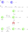
Indirect detection of sulfenic acid. (A) Indirect chemical detection using arsenite reduction. (B) Enzyme activity assay using arsenite mediated reduction of sulfenic acid. (C) Indirect chemical detection using pervanadate mediated hyperoxidation and selective immunoprecipitation.
Fig. 9
Fig. 9
Reaction of NBD-Cl with protein nucleophiles. NBD-Cl may react with various biological nucleophiles with each conjugation product having different absorbance maximum.
Fig. 10
Fig. 10
Detection of in-cell protein sulfenylation with genetically encoded Yap1-cCRD probe. Cells expressing Yap1-cCRD are exposed to peroxide and protein conjugates are extracted with trichloroacetic acid (TCEP). Free thiols are capped by alkylation with iodoacetamide (IAA). Yap1-cCRD-captured proteins are affinity enriched and eluted with reducing agents (DTT or TCEP). After sample enrichment and protease digestion, the resulting peptides are analyzed and identified by LC–MS/MS.
Fig. 11
Fig. 11
Detection of sulfenic acid by chemical reaction. (A) Nucleophilic reaction of dicoordinated sulfur. (B) Reaction of dimedone with sulfenic acid.
Fig. 12
Fig. 12
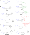
1,3-Dione based probes directly conjugated to biotin or fluorescent tags.
Fig. 13
Fig. 13
(A) Sulfenic acid specific probes with azide or alkyne handles. (B) Enrichment techniques. (C) Alkyne-biotin hapten. (D) Azide-biotin hapten. (E) General in vivo or in vitro protein-SOH labeling scheme.
Fig. 14
Fig. 14
General strategy for labeling and enrichment of sulfenic acid-modified protein and peptides. (A) Direct detection of sulfenylated proteins and peptides using modified TFA-cleavable biotin carbamate linker. (B) Direct detection of sulfenylated proteins with a β-ketoester probe amenable to cleavage by the reaction with hydroxylamine.
Fig. 15
Fig. 15
Quantification of protein sulfenylation using isotope-coded dimedone and iododimedone (ICDID).
Fig. 16
Fig. 16
(A) Anti-dimedone antibody based detection of the cysteine-dimedone protein adducts. (B) Redox-based probes with affinity-based binding module for detecting reversible PTP oxidation.
Fig. 17
Fig. 17
Active-site of Cdc25B showing sulfenic acid mediated disulfide formation between redox-active Cys473 and back-door Cys426. (A) The native Cdc25B (PDB ID: 1QB0) is shown with redox-active Cys473 and back-door Cys426. (B) Sulfenic acid formation at redox-active Cys473 (PDB ID: 1YML). (C) Disulfide formation between redox-active Cys473 and back-door Cys426 (PDB ID: 1CWR).
Fig. 18
Fig. 18
X-ray crystal structures of DJ-1. (A) Wild-type DJ-1 in reduced form (PDB ID: 1P5F); (B) wild type DJ-1 sulfinic acid showing the E18-Cys106 hydrogen bonding (PDB ID: 1SOA); (C) E18D DJ-1 mutant with stable Cys106 sulfenate, hydrogen bonded to D18 (PDB ID: 3CZA).
Fig. 19
Fig. 19
Sulfenic acid metabolism pathways.
Fig. 20
Fig. 20
Kinetic representation of competitive in vivo trapping of sulfenic acid with dimedone in the presence of glutathione.
Fig. 21
Fig. 21
Redox regulation of sulfenic acid—steady state approximation to estimate sulfenic acid concentration.
Scheme 1
Scheme 1
Generation of transient sulfenic acid—nature’s perspective.
Scheme 2
Scheme 2
(1) Fundamental synthetic strategy. (2) General syntheses of transient sulfenic acids.
Scheme 3
Scheme 3
Sulfenic acid mediated generation of 3-isothiazolidinone heterocycle which serves as a chemical model to understand the redox-regulation of PTP1B activity.
Scheme 4
Scheme 4
Synthesis of stable sulfenic acids.
Scheme 5
Scheme 5
Synthesis and reactivity of sulfenic acid embedded in bowl-type cyclophane. (1) Synthesis of Bmt-SOH from Bmt-SH by a mild oxidant. (2) Reactions of Bmt-SOH.

References

    1. Miseta A, Csutora P. Relationship between the occurrence of cysteine in proteins and the complexity of organisms. Mol Biol Evol. 2000;17:1232–1239. - PubMed
    1. Pe’er I, Felder CE, Man O, Silman I, Sussman JL, Beckmann JS. Proteomic signatures: amino acid and oligopeptide compositions differentiate among phyla. Proteins Struct Funct Bioinform. 2004;54:20–40. - PubMed
    1. Sanchez R, Riddle M, Woo J, Momand J. Prediction of reversibly oxidized protein cysteine thiols using protein structure properties. Protein Sci. 2008;17:473–481. - PMC - PubMed
    1. Finkel T. Oxidant signals and oxidative stress. Curr Opin Cell Biol. 2003;15:247–254. - PubMed
    1. Poole LB, Nelson KJ. Discovering mechanisms of signaling-mediated cysteine oxidation. Curr Opin Chem Biol. 2008;12:18–24. - PMC - PubMed

Publication types