

Discrepancy between fluorescence correlation spectroscopy and fluorescence recovery after photobleaching diffusion measurements of G-protein-coupled receptors
- PMID: 23748145
- PMCID: PMC3770895
- DOI: 10.1016/j.ab.2013.04.033
Discrepancy between fluorescence correlation spectroscopy and fluorescence recovery after photobleaching diffusion measurements of G-protein-coupled receptors
Abstract
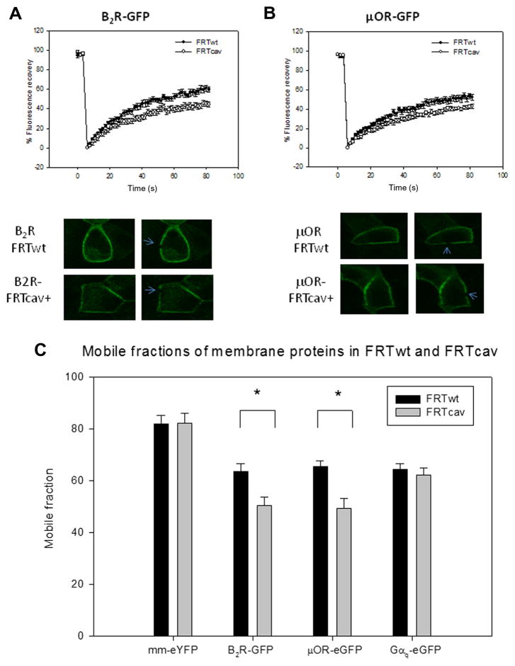
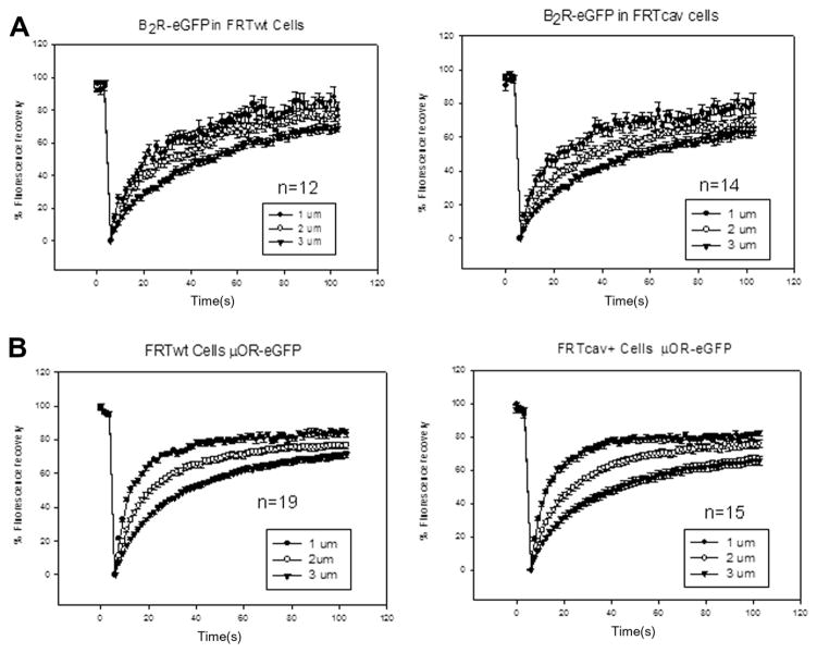
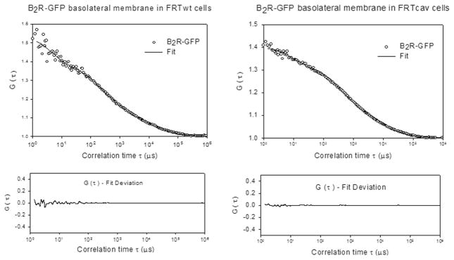
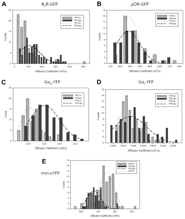
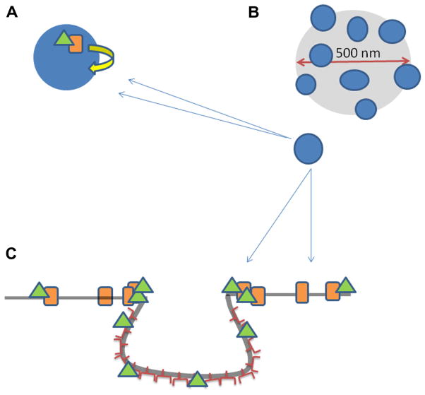
Fluorescence recovery after photobleaching (FRAP) and fluorescence correlation spectroscopy (FCS) are the two most direct methods to measure the diffusion of molecules in intact living cells. Ideally, these methods should produce similar results for an identical system. We have used these methods to monitor the diffusion of two G-protein-coupled receptors and their associated proteins in the plasma membranes of cells that do not or do contain invaginated protein domains called caveolae. FRAP studies show that caveolae domains increase the immobile fraction of receptors without significantly changing their mobility. On the other hand, FCS studies show an unexpected increase the mobility of caveolae-associated proteins. Our data suggest that the geometry of caveolae domains gives rise to a confined diffusion of its attached proteins, resulting in an apparent increase in mobility.
Keywords: Caveolae; Fluorescence correlation spectroscopy; Fluorescence recovery after photobleaching; G-protein-coupled receptors; Protein diffusion.
Copyright © 2013 Elsevier Inc. All rights reserved.
Figures
Similar articles
-
EGF Receptor Stalls upon Activation as Evidenced by Complementary Fluorescence Correlation Spectroscopy and Fluorescence Recovery after Photobleaching Measurements.Int J Mol Sci. 2019 Jul 9;20(13):3370. doi: 10.3390/ijms20133370. Int J Mol Sci. 2019. PMID: 31323980 Free PMC article.
-
Cross-validating FRAP and FCS to quantify the impact of photobleaching on in vivo binding estimates.Biophys J. 2010 Nov 3;99(9):3093-101. doi: 10.1016/j.bpj.2010.08.059. Biophys J. 2010. PMID: 21044608 Free PMC article.
-
Fluorescence Correlation Spectroscopy and Fluorescence Recovery After Photobleaching to study receptor kinase mobility in planta.Methods Mol Biol. 2011;779:225-42. doi: 10.1007/978-1-61779-264-9_13. Methods Mol Biol. 2011. PMID: 21837570
-
Methods to measure the lateral diffusion of membrane lipids and proteins.Methods. 2006 Jun;39(2):147-53. doi: 10.1016/j.ymeth.2006.05.008. Methods. 2006. PMID: 16846741 Review.
-
Recent applications of fluorescence recovery after photobleaching (FRAP) to membrane bio-macromolecules.Sensors (Basel). 2010;10(6):5927-48. doi: 10.3390/s100605927. Epub 2010 Jun 10. Sensors (Basel). 2010. PMID: 22219695 Free PMC article. Review.
Cited by
-
On the Equivalence of FCS and FRAP: Simultaneous Lipid Membrane Measurements.Biophys J. 2016 Jul 12;111(1):152-61. doi: 10.1016/j.bpj.2016.06.001. Biophys J. 2016. PMID: 27410743 Free PMC article.
-
Temporal dependence of shifts in mu opioid receptor mobility at the cell surface after agonist binding observed by single-particle tracking.Sci Rep. 2019 May 13;9(1):7297. doi: 10.1038/s41598-019-43657-x. Sci Rep. 2019. PMID: 31086197 Free PMC article.
-
Super-resolution Visualization of Caveola Deformation in Response to Osmotic Stress.J Biol Chem. 2017 Mar 3;292(9):3779-3788. doi: 10.1074/jbc.M116.768499. Epub 2017 Jan 17. J Biol Chem. 2017. PMID: 28096469 Free PMC article.
-
Cell shape information is transduced through tension-independent mechanisms.Nat Commun. 2017 Dec 15;8(1):2145. doi: 10.1038/s41467-017-02218-4. Nat Commun. 2017. PMID: 29247198 Free PMC article.
-
Heterologous regulation of Mu-opioid (MOP) receptor mobility in the membrane of SH-SY5Y cells.J Biol Chem. 2014 Oct 10;289(41):28697-706. doi: 10.1074/jbc.M114.588558. Epub 2014 Sep 2. J Biol Chem. 2014. PMID: 25183007 Free PMC article.
References
Publication types
MeSH terms
Substances
Grants and funding
LinkOut - more resources
Full Text Sources
Other Literature Sources
Miscellaneous
