Review
. 2013 Sep:137:136-49.
doi: 10.1016/j.jsbmb.2013.05.012. Epub 2013 Jun 6.

AKR1C3 as a target in castrate resistant prostate cancer

Affiliations
Review

AKR1C3 as a target in castrate resistant prostate cancer

Adegoke O Adeniji et al. J Steroid Biochem Mol Biol. 2013 Sep.

Abstract

Aberrant androgen receptor (AR) activation is the major driver of castrate resistant prostate cancer (CRPC). CRPC is ultimately fatal and more therapeutic agents are needed to treat this disease. Compounds that target the androgen axis by inhibiting androgen biosynthesis and or AR signaling are potential candidates for use in CRPC treatment and are currently being pursued aggressively. Aldo-keto reductase 1C3 (AKR1C3) plays a pivotal role in androgen biosynthesis within the prostate. It catalyzes the 17-ketoreduction of weak androgen precursors to give testosterone and 5α-dihydrotestosterone. AKR1C3 expression and activity has been implicated in the development of CRPC, making it a rational target. Selective inhibition of AKR1C3 will be important, however, due to the presence of closely related isoforms, AKR1C1 and AKR1C2 that are also involved in androgen inactivation. We examine the evidence that supports the vital role of AKR1C3 in CRPC and recent developments in the discovery of potent and selective AKR1C3 inhibitors. This article is part of a Special Issue entitled 'CSR 2013'.

Keywords: Androgens; Nonsteroidal anti-inflammatory drugs; Prostaglandin F synthase; Prostate cancer; Type 5 17β-hydroxysteroid dehydrogenase.


Figures

Figure 1
Figure 1
AKR1C3 and Androgen Metabolism in The Prostate (Δ5-Adiol, 5-Androstene-3β,17β-diol; Δ4-Adione, 4-Androstene-3,17-dione; 5α-Adione, 5α-Androstane-3,17-dione; AR, Androgen receptor; ARE, Androgen response element; DHEA, Dehydroepiandrosterone; 5α-DHT, 5α-Dihydrotestosterone; HSD3B, 3β-Hydroxysteroid dehydrogenase; PREG, Pregnenolone; SRD5A, 5α-Reductase); enzymes are also listed as their gene names.
Figure 2
Figure 2
AKR1C3 and Prostaglandin Synthesis
Figure 3
Figure 3
Structure and Potency of AKR1C3 Inhibitors. Rings are designated A and B for ease of discussion in the text. FLU = flufenamic acid.
Figure 4
Figure 4
Representative Structures of Indomethacin based AKR1C3 Inhibitors. Compound 13 (n=2): R1 = OCH3, R2 = OH, R3 = CH3, R4 = Cl, AKR1C3 IC50 = 0.22 μM AKR1C2 IC50 = 56.5 μM; compound 14 (n=1): R1 = OCH3, R2 = OH and R3 = Cl, AKR1C3 IC50 = 0.96 μM, AKR1C2 IC50 = 100 μM; compound 15 (n=1): R1 = OCH3, R2 = CH2CH3, R3, = H, R4 = OH, R5 = Cl AKR1C3 IC50 = 0.09 μM AKR1C2 IC50 = 49.6 μM..
Figure 5
Figure 5
Ligand binding pocket of AKR1C3 and AKR1C2. AKR1C3 and AKR1C2 share very similar overall structures. (A), sideview of the overall structure of AKR1C3 in complex with natural substrate prostaglandin D2 (PGD2, blue). (B), topview of the overall structure of AKR1C2 in complex with testosterone (green). Cofactor NADP+ is colored in red. β-strands are colored in yellow. The surface of the subpockets defined by Byrns et al. [54] are shown in the cross-section view of the ligand binding site of AKR1C3 (C) and AKR1C2 (D). AKR1C3 has much bigger subpockets compared to AKR1C2. The ligands and cofactors are not on the same plane as the protein structure/surface. The surface is colored according to protein secondary structure: yellow, β-stands; gray, α-helices and flexible loops. SC, steroid channel; OS, oxyanion site; SP, subpocket. All figures are prepared using The PyMOL Molecular Graphics System, Version 1.20 Schrödinger, LLC.
Figure 6
Figure 6
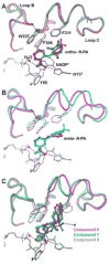
Structures of the N-PA based analogs and 3-(3,4-dihydroisoquinolin-2(1H)-ylsulfonyl)benzoic acids bound to AKR1C3. Both classes of inhibitors utilize the oxyanion hole - SP1 cavity. Two slightly different binding poses exist for the N-PAs depending on the position of the carboxylate. A, for the o-CO2H-substituted N-PAs compounds, FLU (red), meclofenamic acid (blue), and mefenamic acid (green), the amine bridge forms hydrogen bonds with the cofactor nicotinamide head. B, for the m-CO2H-substituted N-PA based analogs, compound 1 (cyan) and 3-PBA (magenta), the bridge atom moves 2.4 Å away from the cofactor and no longer maintains the interaction. Compared to FLU, compound 1 projects 1 Å deeper into the SP1 pocket and induces significant movement of Phe306 and Phe311, which could be basis for its selectivity. C, Structures of the prototypical 3-(3,4-dihydroisoquinolin-2(1H)-ylsulfonyl)benzoic acids, compounds 6 (magenta), 7 (green), and 8 (gray). Even though there is only a slight change in the binding position between compounds 6 and 7, Trp227 is flipped and the C-terminal loop (Loop C: Asp300-end) is shifted to accommodate the two compounds.The presence of the methylamide in compound 8 causes significant movements of the benzyl ring and the sulfonyl linker. But the tetrahydroquinoline group is still positioned in the SP1 pocket. The linker regions of the N-PAs and the 3-(3,4-dihydroisoquinolin-2(1H)-ylsulfonyl)benzoic acids occupy the same location and the 3-(3,4-dihydroisoquinolin-2(1H)-ylsulfonyl)benzoic acids reach the same depth in the SP1 pocket as compound 1. But the plane of the bicyclic ring is roughly perpendicular to the phenyl ring in the N-PA compounds. Hydrogen bonds are shown as red dashes. Only the residues that are normally involved in significant movement are shown. Water molecules are shown as spheres. Distance is indicated by blue dashes.
Figure 7
Figure 7
Structures of indomethacin analogs bound to AKR1C3. A, overlay of the two indomethacin conformations at different pH values. The pH 6.0 conformation (yellow) utilizes the SP3 pocket, whereas the pH 7.5 conformation (red) occupies the oxyanion site and the SP1 pocket. At intermediate pH (pH 6.8, PDB ID 3UGR)[77], the crystal structure shows presence of both conformations (70% occupancy by the low pH conformation, 25% by the high pH conformation, with the remaining 5% unaccounted for). Hydrogen bonds for the pH 6.0 conformation are shown as red dashes. B, two molecules of 2′-des-methyl-indomethacin (blue) are bound per active site. Only the lower molecule closer to the oxyanion site is contributing to inhibition. The accommodation of the second molecule forces significant loop movements of AKR1C3 compared to other indomethacin analogs. (indomethacin, yellow; sulindac, magenta) structures. C, overlay of indomethacin pH 7.5 conformation and zomepirac (green). Zomepirac adopts a very similar conformation to indomethacin. D, sulindac (magenta) is anchored to the oxyanion site but binds quite differently from the indomethacin conformation by extending towards the SP2 pocket.
Figure 8
Figure 8
Structures of the arylpropionic acids bound to AKR1C3. A, overlay of (R)-flurbiprofen (blue) and (R)-ibuprofen (yellow) bound to AKR1C3. The two compounds adopt very similar binding poses despite their structural difference. B, overlay of (R)-naproxen (green) and (S)-naproxen (red). AKR1C3 shows only subtle preference for the (R)-configuration, which is reflected by the limited changes in binding conformation imposed by the chirality of the compounds. Hydrogen bonds are shown as red dashes.
Figure 9
Figure 9
Structure of the bifunctional analog compound 10 (magenta) shown in overlay with compounds 1 (cyan) bound to AKR1C3. Two molecules of compound 10 are bound per AKR1C3 active site and the first molecule resembles the binding conformation of the N-PA compounds. The naphthyl rings of compound 10 are perpendicular to the B-ring of compound 1.
Figure 10
Figure 10
Tiered Approach to Preclinical Development of AKR1C3 Inhibitors

References

    1. SEER Cancer Statistics Review, 1975–2009. National Cancer Institute; Bethesda, MD: 2010. based on November 2009 SEER data submission, posted to the SEER web site, 2010. http://seer.cancer.gov/statfacts/html/prost.html.
    1. Jemal A, Siegel R, Xu J, Ward E. Cancer statistics, 2010. CA Cancer J Clin. 2010;60(5):277–300. - PubMed
    1. http://www.cancer.org/Cancer/ProstateCancer/DetailedGuide/prostate-cancer-key-statistics.

    1. Knudsen KE, Scher HI. Starving the addiction: new opportunities for durable suppression of AR signaling in prostate cancer. Clin Cancer Res. 2009;15(15):4792–4798. - PMC - PubMed
    1. Knudsen KE, Penning TM. Partners in crime: deregulation of AR activity and androgen synthesis in prostate cancer. Trends Endocrinol Metab. 2010;21(5):315–324. - PMC - PubMed

Publication types

MeSH terms

Substances