. 2014 Aug;9(8):1134-42.
doi: 10.1093/scan/nst096. Epub 2013 Jun 6.

Neural substrates of classically conditioned fear-generalization in humans: a parametric fMRI study

Affiliations

Neural substrates of classically conditioned fear-generalization in humans: a parametric fMRI study

Shmuel Lissek et al. Soc Cogn Affect Neurosci. 2014 Aug.

Abstract

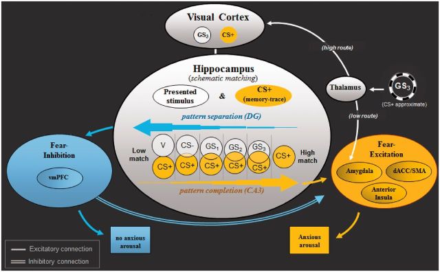
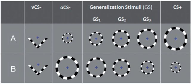
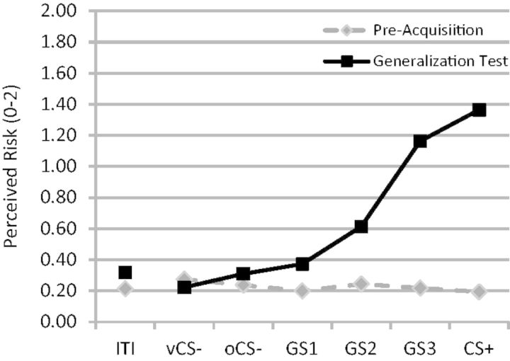
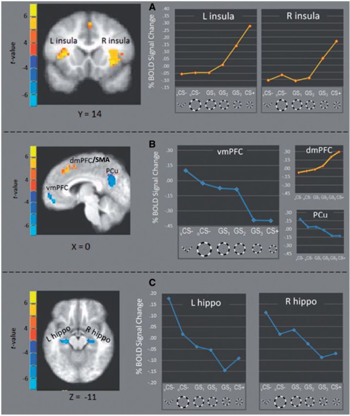
Recent research on classical fear-conditioning in the anxiety disorders has identified overgeneralization of conditioned fear as an important conditioning correlate of anxiety pathology. Unfortunately, only one human neuroimaging study of classically conditioned fear generalization has been conducted, and the neural substrates of this clinically germane process remain largely unknown. The current generalization study employs a clinically validated generalization gradient paradigm, modified for the fMRI environment, to identify neural substrates of classically conditioned generalization that may function aberrantly in clinical anxiety. Stimuli include five rings of gradually increasing size with extreme sizes serving as cues of conditioned danger (CS+) and safety (CS-). The three intermediately sized rings serve as generalization stimuli (GSs) and create a continuum-of-size from CS+ to CS-. Results demonstrate 'positive' generalization gradients, reflected by declines in responding as the presented stimulus differentiates from CS+, in bilateral anterior insula, dorsomedial prefrontal cortex, and bilateral inferior parietal lobule. Conversely, 'negative' gradients, reflected by inclines in responding as the presented stimulus differentiates from CS+ were instantiated in bilateral ventral hippocampus, ventromedial prefrontal cortex and precuneus cortex. These results as well as those from connectivity analyses are discussed in relation to a working neurobiology of conditioned generalization centered on the hippocampus.

Keywords: anxiety; conditioned generalization; fMRI; fear-conditioning; neurobiology.


Figures

Fig. 1
Fig. 1
Neural model of conditioned fear generalization. Following acquisition of fear to CS+, when exposed to a stimulus resembling CS+ (i.e. GS 3 ), the thalamus is thought to relay sensory information about GS 3 to amygdala-based fear circuits via a ‘quick and dirty’ route resulting in a fast initial fear response to GS 3 . The thalamus simultaneously sends sensory GS 3 information to visual cortices for higher level sensory processing—a slower route through which neural representations of GS 3 are activated in visual cortex. Next, through hippocampally based ‘schematic matching’, the overlap between patterns of brain activity representing GS 3 and the previously encoded CS+ is assessed. Given sufficient overlap, CA3 neurons in hippocampus are thought to initiate ‘pattern completion’ (e.g. Treves and Rolls, 1994 ), whereby a subset of cues from a previous experience (i.e. CS+) activates the stored pattern representing that experience. Pattern completion by the hippocampus is then proposed to result in activation of brain structures associated with fear excitation (denoted in yellow: anterior insula, dACC, amygdala), culminating in the autonomic, neuroendocrine and behavioral constituents of the fear response. In the event of insufficient overlap between neural representations of GS 3 and the CS+, dentate gyrus neurons in the hippocampus are thought to initiate ‘pattern separation’ (e.g. McHugh et al. , 2007 ), resulting in the spread of activation to structures associated with fear inhibition (denoted in blue: vmPFC). Such activations are then proposed to attenuate ongoing activity in amygdala-based fear circuits initiated earlier by the ‘quick and dirty’ route and serve to stem anxious arousal. GS 1 , GS 2 , GS 3 = ring-shaped generalization stimuli; CS+ = ring-shaped danger-cue; CS− = ring shaped safety cue; vCS− = V-shaped safety cue; DG = dentate gyrus; CA3 = cornu ammonis region 3; vmPFC = ventromedial prefrontal cortex; dACC = dorsal anterior cingulate cortex; SMA = supplementary motor cortex area.
Fig. 2
Fig. 2
Conditioning and generalization stimuli for counterbalancing groups A and B. Half of the participants were assigned to counterbalancing group A and half to B. For both counterbalancing groups A and B , GS 3 consisted of the ring closest in size to CS+, with GS 2 and GS 1 further decreasing in similarity to CS+. Ring diameters in centimetres (and visual angles) from smallest to largest were: 6.63 (0.93°), 8.02 (1.12°), 9.38 (1.31°), 10.98 (1.54°) and 12.46 (1.75°). vCS− = v-shaped conditioned safety cue; oCS− = ring-shaped conditioned safety cue; GS 1 , GS 2 and GS 3 = three classes of generalization stimuli; CS+ = conditioned danger cue.
Fig. 3
Fig. 3
Online ratings of shock risk (0 = no risk, 1 = some risk, 2 = high risk) both before acquisition training (pre-acquisition) and after (generalization test). ITI = inter-trial interval; vCS− = v-shaped conditioned safety cue; oCS− = ring-shaped conditioned safety cue; GS 1 , GS 2 and GS 3 = three classes of generalization stimuli; CS+ = conditioned danger cue.
Fig. 4
Fig. 4
fROIs responding more strongly to CS+ vs CS− (yellow) and CS− versus CS+ (blue) fell along positive and negative generalization gradients, respectively. Specifically, activations in left and right anterior insula ( A ), as well as dmPFC ( B ), fell along positive gradients with increasing signal change as presented stimuli became more similar to CS+. Conversely, activations in vmPFC, and PCC (B) as well as left and right anterior hippocampus ( C ) formed negative gradients with decreasing levels as presented stimuli became more similar to CS+. L = left; R = right; vmPFC = ventromedial prefrontal cortex; dmPFC = dorsomedial prefrontal cortex; SMA = supplementary motor cortex; Pcu = precuneus; hippo = hippocampus.

References

    1. Alvarez RP, Biggs A, Chen G, Pine DS, Grillon C. Contextual fear conditioning in humans: cortical-hippocampal and amygdala contributions. Journal of Neuroscience. 2008;28:6211–9. - PMC - PubMed
    1. Antunes R, Moita MA. Discriminative auditory fear learning requires both tuned and nontuned auditory pathways to the amygdala. Journal of Neuroscience. 2010;30:9782–7. - PMC - PubMed
    1. Armony JL, Servan-Schreiber D, Romanski LM, Cohen JD, LeDoux JE. Stimulus generalization of fear responses: effects of auditory cortex lesions in a computational model and in rats. Cerebral Cortex. 1997;7:157–65. - PubMed
    1. Bucci DJ, Saddoris MP, Burwell RD. Contextual fear discrimination is impaired by damage to the postrhinal or perirhinal cortex. Behavioral Neuroscience. 2002;116:479–88. - PubMed
    1. Büchel C, Morris J, Dolan RJ, Friston KJ. Brain systems mediating aversive conditioning: an event-related fMRI study. Neuron. 1998;20:947–57. - PubMed

Publication types