

Colony forming unit endothelial cells do not exhibit telomerase alternative splicing variants and activity
- PMID: 23748893
- PMCID: PMC3770257
- DOI: 10.6091/ibj.1100.2013
Colony forming unit endothelial cells do not exhibit telomerase alternative splicing variants and activity
Abstract
Introduction: Endothelial progenitor colony forming unit-endothelial cells (CFU-EC) were first believed to be the progenitors of endothelial cells, named endothelial progenitor cells. Further studies revealed that they are monocytes regulating vasculogenesis. The main hindrance of these cells for therapeutic purposes is their low frequency and limited replicative potentials. This study was undertaken to determine telomerase activity and alternative splicing variants in CFU-EC as a potential cause of limited replicative capacity in these cells.
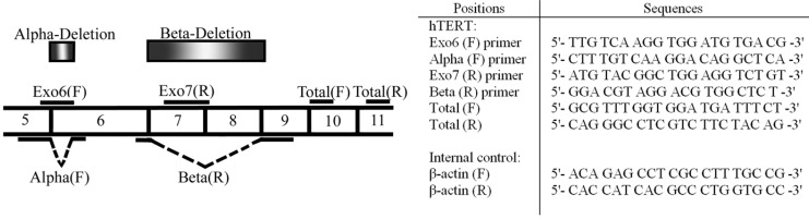
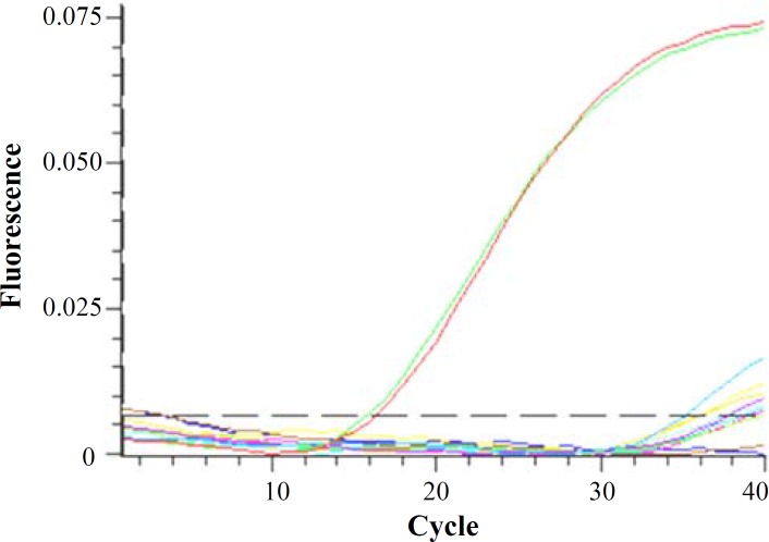
Methods: CFU-EC were isolated from peripheral blood using a standard cell culture assay. Colonies were detached mechanically and alternative splicing variant mRNA were evaluated using real-time PCR. Telomerase enzyme activity was assessed using telomerase repeat amplification protocol. The same procedures were done on the cancer cell line Calu6 as the positive control.
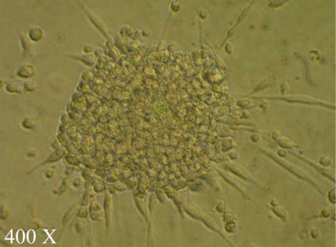
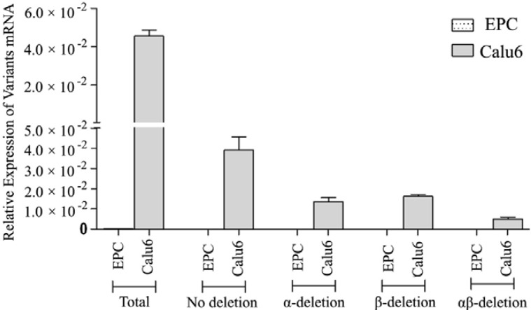
Results: The cultured peripheral blood mononuclear cells formed colonies with spindle-shaped monocytic cells sprouted from the clusters. These morphological characteristics fulfill the definition of CFU-EC. Telomere length amplification protocol assay revealed no telomerase activity and real-time PCR showed no expression of telomerase enzyme mRNA in CFU-EC. Both parameters were significantly higher in the cancer cell line Calu6 taken as the positive control.
Conclusion: The absence of telomerase activity in the CFU-EC is a result of pre-transcriptional regulation of gene expression rather than other mechanisms for controlling telomerase activity such as post-transcriptional modifications. This finding can explain the limited proliferative activity of CFU-EC cells. We propose that absence of telomerase activity in CFU-EC can be attributable to their more mature monocytic nature that needs further investigations.
Keywords: Telomerase; Endothelial cells; Hemangioblast; Alternative splicing.
Figures
References
-
- Shi Q, Rafii S, Wu MH, Wijelath ES, Yu C, Ishida A, et al. Evidence for circulating bone marrow-derived endothelial cells. Blood. 1998 Jul;92(2):362–7. - PubMed
-
- Asahara T, Murohara T, Sullivan A, Silver M, Zee RVD, Li T, et al. Isolation of putative progenitor endothelial cells for angiogenesis. Science. 1997 Feb;275:964–7. - PubMed
-
- Hill JM, Zalos G, Halcox JP, Schenke WH, Waclawiw MA, Quyyumi AA, et al. Circulating endothelial progenitor cells, vascular function, and cardiovascular risk. N Engl J Med. 2003 Feb;348(7):593–600. - PubMed
Publication types
MeSH terms
Substances
LinkOut - more resources
Full Text Sources
Medical