. 2013:4:1978.
doi: 10.1038/ncomms2978.

Haemodynamically dependent valvulogenesis of zebrafish heart is mediated by flow-dependent expression of miR-21

Affiliations
Free PMC article

Haemodynamically dependent valvulogenesis of zebrafish heart is mediated by flow-dependent expression of miR-21

Toshihiro Banjo et al. Nat Commun. 2013.
Free PMC article

Abstract

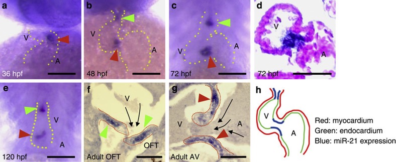
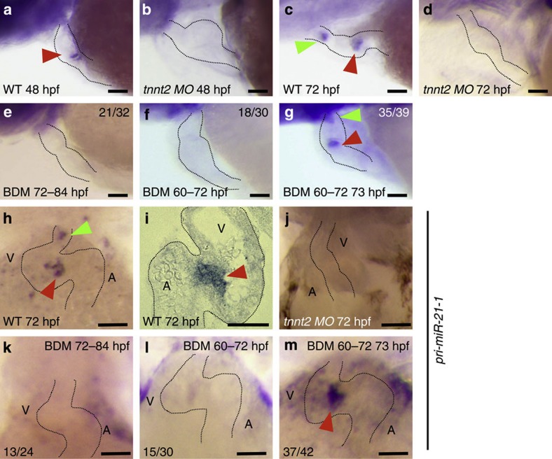
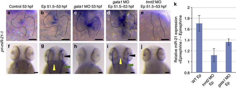
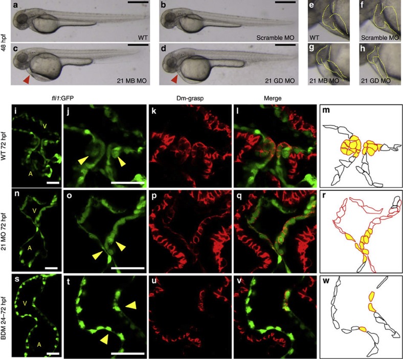
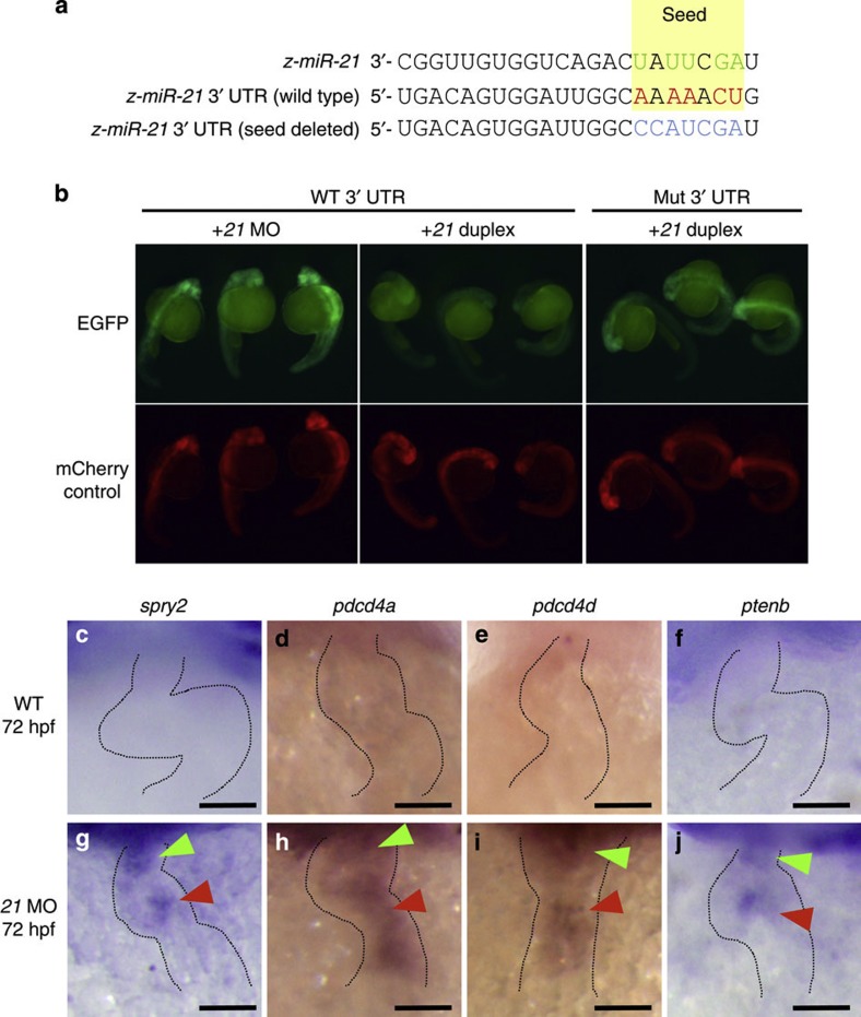
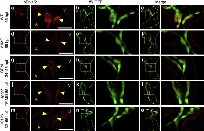
Heartbeat is required for normal development of the heart, and perturbation of intracardiac flow leads to morphological defects resembling congenital heart diseases. These observations implicate intracardiac haemodynamics in cardiogenesis, but the signalling cascades connecting physical forces, gene expression and morphogenesis are largely unknown. Here we use a zebrafish model to show that the microRNA, miR-21, is crucial for regulation of heart valve formation. Expression of miR-21 is rapidly switched on and off by blood flow. Vasoconstriction and increasing shear stress induce ectopic expression of miR-21 in the head vasculature and heart. Flow-dependent expression of mir-21 governs valvulogenesis by regulating the expression of the same targets as mouse/human miR-21 (sprouty, pdcd4, ptenb) and induces cell proliferation in the valve-forming endocardium at constrictions in the heart tube where shear stress is highest. We conclude that miR-21 is a central component of a flow-controlled mechanotransduction system in a physicogenetic regulatory loop.


Figures

Figure 1
Figure 1. Expression patterns of miR-21.
(a) Soon after initiation of blood flow (36 hpf), miR-21 began to be expressed in a subset of the endocardial cells in the AV canal (red arrowhead). A, atrium; V, ventricle. (b) At 48 hpf, endocardial expression in the OFT became evident (green arrowhead). Expression at the AV canal was arranged around the canal lumen (red arrowhead). (c,d) The same expression patterns were maintained at 72 hpf (c) and 120 hpf (e). (d) In sections, miR-21 expression was detected only in the endocardial cells, not in the myocardial layer. (f,g) Expression of miR-21 was detected in the valves of adult fish at the OFT (green arrowheads in f) and the AV canal (red arrowheads in g). Dotted red lines indicate contours of the valves. Black arrows indicate the direction of blood flow. (h) A schematic representation of miR-21 expression only in specific regions of the endocardium of the OFT and the AV canal (blue lines). Scale bars, 50 μm.
Figure 2
Figure 2. Effect of heartbeat on expression of miR-21 in the developing heart tube.
(a,c) Normal expression of miR-21 at the OFT (green arrowhead) and the AV canal (red arrowheads) at 48 (a) and 72 hpf (c). Dotted lines outline the heart. (b,d). In the tnnt2 morphants, expression of miR-21 was detected neither at 48 (b) nor at 72 hpf (d). (eg) Twelve hours arrest of heartbeat (from 72 to 84 hpf in e, and from 60 to 72 hpf in f) obliterated the two patches of miR-21 expression, whereas just 1 h of heartbeat restored the full expression (g). (hm) pri-miR-21-1 transcripts were expressed in the same patterns at 72 hpf (whole mount in h, and section in i). This expression was lost in the tnnt2 morphant heart (j). Expression of pri-miR-21 disappeared after 12 h cardiac arrest (from 72 to 84 hpf in k, and from 60 to 72 hpf in l) and 1 h of heartbeat restored it (red arrowhead in m). WT, wild type; MO, morphant; BDM, treated with BDM. Scale bars, 50 μm.
Figure 3
Figure 3. Expression patterns of pri-miR-21-1, the primary transcript of miR-21.
Expression of pri-miR-21-1 in the heart (ae) and the head vasculature (fj) at 53 hpf. The WT (a,b,f and g), gata1 morphants (c,d,h and i) and tnnt2 morphants (e,j) were stimulated by epinephrine (Epi) from 51.5 to 53 hpf (b,d,e,g,i and j). Without Epi stimulation, pri-miR-21-1 was expressed only in the valve-forming endocardium (a), not in the head vasculature (f). Elimination of blood cells from the circulation by knocking down gata1 did not affect the expression (c,h). After stimulation by Epi, expression of pri-miR-21-1 became slightly stronger at the AV canal (b). (g) Expression in the head became evident in the metencephalic artery/middle cerebral vein (yellow arrowheads), the primary head sinus (green arrowheads) and the primordial mid-brain channel (black arrowheads). This ectopic induction was not affected in the gata1 morphants (i), but the absence of heartbeat cancelled it (j). (k) Relative expression of the mature miR-21 was measured by qRT–PCR using RNA isolated from the whole bodies. In the WT, Epi induced ~1.7-fold, whereas the induction in the tnnt2 morphant was minimal. In the gata1 morphants, miR-21 was induced at a relatively lower extent than the WT (~1.4-fold). Scale bars, 50 μm (ae), 100 μm (fj).
Figure 4
Figure 4. A valveless miR-21 morphant.
(ad) Blood flow of the miR-21 morphants stagnated with pooling of blood cells on the yolk at 48 hpf (red arrowhead in c,d) (MB, multiblocking MO; GD, Guide Dicer MO). This stagnation was neither present in the WT (a) nor embryos injected with a scrambled MO (b). Higher magnification views are shown in eh, with heart contours indicated by dotted lines. (im) At 72 hpf, thick layers of the GFP-positive endocardial cells were protruded into the AV canal (i), with cuboidal thickening cells (yellow arrowheads in j). Cell shapes are visualized by Dm-grasp staining (k,l). Dm-grasp- and GFP-positive cells are cuboidal, whereas Dm-grasp-negative and GFP-positive cells are squamous (l). (m) A schematic representation of the endocardial cells, with cells in the valve-forming area shown in yellow, Dm-grasp-positive cells in red lines, negative cells in black lines, respectively. A, atrium, V, ventricle. (nr) In the miR-21 morphants, only a thin layer of endocardial cells was evident with neither thickening nor protrusion into the lumen. (sw) When heartbeat was stopped from 24 to 72 hpf by BDM, neither thickening nor protrusion of the endocardium was initiated. The number of the Dm-grasp-positive endocardial cells was reduced. Scale bars, 500 μm (ad), 50 μm (i,j,n,o,s,t).
Figure 5
Figure 5. Sequence alignment and expression patterns of selected genes.
(a) Sequence alignment of zebrafish miR-21, 3′-UTR of spry2 and its mutated version is shown, with the seed sequences highlighted in yellow and the complementary nucleotides in red and green. (b) The EGFP-spry2 reporter with the WT spry2 3′-UTR gave strong fluorescent signals when the endogenous miR-21 was blocked by the MO against it (left panel), but failed to show the signals when co-injected with the miR-21 duplex (middle panel). The EGFP-spry2 reporter mRNA, in which the seed sequence was mutated, produced strong signals even when co-injected with the miR-21 duplex (right panel) at 24 hpf. (c,g) Expression of the endogenous spry2 was not observed in the WT at 72 hpf (c), whereas it was induced in both the OFT and AV canal in the morphant (green and red arrowheads in g, respectively). (dj) Other target genes, pdcd4a (d,h), pdcd4b (e,i) and ptenb (f,j) were induced in the morphant (hj). Expression of these genes was not observed in the WT (cf). Scale bars, 50 μm (cj).
Figure 6
Figure 6. Effects of a gain of function of spry2 and U0126 treatment.
(ah) When the MAP kinase cascade was blocked by injection of a target protection (TP) MO of spry2 (b,f) or U0126-treatment (d,h), hearts (dotted outlines) exhibited incomplete looping (f,h). Red arrowhead indicates pooling of blood, a sign of stagnation of circulation. The normal looped heart is shown in (e). (i–w) As observed in the miR-21 morphants and the arrested hearts, the endocardial cells were single-layered, flattened and stretched in the hearts injected with the TP MO of spry2 (nr) and the U0126-treated embryos (sw). Normal thickening of cells observed in the WT (im) was lost, as visualized by fli1-GFP reporter and Dm-grasp staining. As illustrated in a schematic representation (m, r and w), the multi-layered cuboidal cells observed in the WT (m) was detected neither in the hearts injected with the target protection MO of spry2 (r) or treated by U0126 (w). Scale bars, 500 μm (ad), 50 μm (i,j,n,o,s,t).
Figure 7
Figure 7. Suppression of MAP kinase signalling in miR-21 morphants and BDM-treated hearts.
(ac) In the WT beating hearts, a phosphorylated form of Erk1/2 (pErk1/2) was stained in the cuboidal endocardial cells at the AV junction (yellow arrowheads in a). The entire endocardial cells were labelled by GFP, which expression was driven by the fli1 promoter (b,c). (df) In the miR-21 morphants, the pErk1/2 staining was week and the cells were flat. (gi) When the heartbeat was stopped by BDM, phosphorylation of Erk1/2 was also suppressed, leaving only faint staining in the apical surface of flattened endocardial cells. (jo) Injection of the miR-21 target protection MO and the U0126 treatment gave the same results on pErk1/s staining. A, atrium; V, ventricle. Scale bars, 20 μm.

References

    1. Srivastava D. Making or breaking the heart: from lineage determination to morphogenesis. Cell 126, 1037–1048 (2006). - PubMed
    1. Stainier D. Y. Zebrafish genetics and vertebrate heart formation. Nat. Rev. Genet. 2, 39–48 (2001). - PubMed
    1. Forouhar A. S. et al. The embryonic vertebrate heart tube is a dynamic suction pump. Science 312, 751–753 (2006). - PubMed
    1. Hove J. R. et al. Intracardiac fluid forces are an essential epigenetic factor for embryonic cardiogenesis. Nature 421, 172–177 (2003). - PubMed
    1. Combs M. D. & Yutzey K. E. Heart valve development: regulatory networks in development and disease. Circ. Res. 105, 408–421 (2009). - PMC - PubMed

Publication types

MeSH terms