Review
. 2014 Mar;71(5):793-811.
doi: 10.1007/s00018-013-1380-7. Epub 2013 Jun 11.

The blockade of the neurotransmitter release apparatus by botulinum neurotoxins

Affiliations
Review

The blockade of the neurotransmitter release apparatus by botulinum neurotoxins

Sergio Pantano et al. Cell Mol Life Sci. 2014 Mar.

Abstract

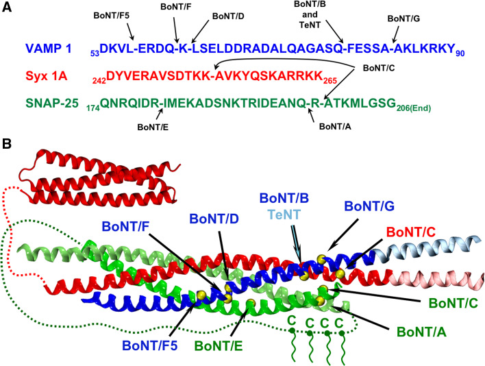
The high toxicity of the seven serotypes of botulinum neurotoxins (BoNT/A to G), together with their specificity and reversibility, includes them in the list A of potential bioterrorism weapons and, at the same time, among the therapeutics of choice for a variety of human syndromes. They invade nerve terminals and cleave specifically the three proteins which form the heterotrimeric SNAP REceptors (SNARE) complex that mediates neurotransmitter release. The BoNT-induced cleavage of the SNARE proteins explains by itself the paralysing activity of the BoNTs because the truncated proteins cannot form the SNARE complex. However, in the case of BoNT/A, the most widely used toxin in therapy, additional factors come into play as it only removes a few residues from the synaptosomal associate protein of 25 kDa C-terminus and this results in a long duration of action. To explain these facts and other experimental data, we present here a model for the assembly of the neuroexocytosis apparatus in which Synaptotagmin and Complexin first assist the zippering of the SNARE complex, and then stabilize and clamp an octameric radial assembly of the SNARE complexes.


Figures

Fig. 1
Fig. 1
Cartoon representation and domain composition of Botulinum Neurotoxins. Comparison between the crystallographic structures of BoNT/A (a) and BoNT/E (b). The L chain, translocation HN, binding HC–N, and HC–C domains are colored in blue, green, yellow, and red, respectively. The Zn2+ atom bound to the active site of the matalloprotease domain is shown in violet. Notice the different orientation of the HC domains of BoNT/E with respect to that of BoNT/A; the global architecture of BoNT/B (not shown) is nearly identical to that of BoNT/A
Fig. 2
Fig. 2
The SNARE complex and the cleavage sites of BoNTs. a Primary sequences of the three SNARE proteins in the neighborhood of the cleavage site of the different BoNTs. Numbering and sequences here and along the manuscript correspond to mouse (Mus musculus) unless otherwise stated. Gaps in the sequences indicate the peptide bond cleaved by each serotype. b Cartoon representation of the SNARE complex. VAMP, Syntaxin, and SNAP25 are shown in blue, red, and green, respectively. Light colors in VAMP and Syntaxin indicate TM segments, which extend beyond the four-helix bundle of the SNARE complex according the PDB structure 3IPD. Red dashed lines indicate unstructured segments between the N-terminal three-helix bundle of Syntaxin (PDB id: 1EZ3) and the SNARE core domain. The green dashed line represents the long linker existing between the two helices of SNAP25, which contains four conserved palmitoylated cysteines. The yellow spheres correspond to the amino acid before the cleavage point of each serotype. This figure is intended only to provide a comprehensive view of the possible impact of each BoNT on the formation of the SNARE complex. Notice, however, that BoNTs do not cleave the SNAREs when they are engaged in the four-helix bundle
Fig. 3
Fig. 3
Multiple sequence alignment of SNARE proteins. Primary sequences of the entire proteins were collected from the UniProt database (http://www.uniprot.org) and aligned using T-COFFEE at http://www.tcoffee.org/. Only relevant regions around the cleavage sites are shown. Positive, negative and polar amino acids are back shaded in blue, red and green, respectively. a The different variants of VAMP correspond to the following UniProt sequence codes: Q62442 (VAMP1 Mouse), P63044 (VAMP2 Mouse), P63024 (VAMP3 Mouse), O70480 (VAMP4 Mouse), P70280 (VAMP4 Mouse), O89116 (VTI1A Mouse); Q63666 (VAMP1 Rat), P63045 (VAMP2 Rat), P63025 (VAMP3 Rat), D4A560 (VAMP4 Rat); P23763 (VAMP1 Human), P63027 (VAMP2 Human), Q15836 (VAMP3 Human), O75379 (VAMP4 Human); F1NJC6 (VAMP1 Chicken), F1NTL8 (VAMP2 Chicken), F1P4I3 (VAMP3 Chicken) and F1NLG6 (VAMP1 Chicken). b The different variants of Syntaxin correspond to the following UniProt sequence codes: O35526 (Syx1A Mouse), P61264 (Syx1B Mouse), Q00262 (Syx2 Mouse), Q64704 (Syx3 Mouse), P70452 (Syx4 Mouse), Q8K1E0 (Syx5 Mouse); P32851 (Syx1A Rat), P61265 (Syx1B Rat), P50279 (Syx2 Rat), Q08849 (Syx3 Rat), Q08850 (Syx4 Rat), Q08851 (Syx5 Rat); Q16623 (Syx1A Human), P61266 (Syx1B Human), P32856 (Syx2 Human), Q13277 (Syx3 Human), Q12846 (Syx4 Human), Q13190 (Syx5 Human); F1N861 (Syx1A Chicken), O42340 (Syx1B Chicken), F1NW46 (Syx2 Chicken), H9KZA6 (Syx3 Chicken) and Q5ZL19 (Syx6 Chicken). c The variants of SNAP25 (SNAP23 and SNAP29) correspond to the following UniProt sequence codes: P60879 (SNAP25 Mouse), O09044 (SNAP23 Mouse), Q9ERB0 (SNAP29 Mouse); P60881 (SNAP25 Rat), O70377 (SNAP23 Rat), Q9Z2P6 (SNAP29 Rat); P60880 (SNAP25 Human), O00161 (SNAP23 Human), O95721 (SNAP29 Human); P60878 (SNAP25 Chicken), E1BRL4 (SNAP23 Chicken) and F1NYA3 (SNAP29 Chicken)
Fig. 4
Fig. 4
Interaction between BoNTs and SNARE proteins. a Cartoon representation of the X-ray structure of BoNT/A in complex with SNAP25 (green). The color in the structure of the enzyme varies from red (near the active site) to white (far from the active site). The Zn2+ atom and the catalytic water bound to the active site are shown as violet and yellow spheres, respectively. The metal coordinating residues His223, 227 and Asn224 (corresponding to Glu224, which is mutated in this structure) are shown in sticks. The second shell coordination residues Arg363 and Phe366 (corresponding to Tyr366, mutated in this structure) are also shown in sticks and indicated with black ovals. b Same as (a) for the complex between BoNT/F and VAMP (blue). The position of the catalytic water was not determined in this structure
Fig. 5
Fig. 5
Molecular representation of Synaptotagmin and CPX. a Cartoon representation of the Ca2+ bound state of Synaptotagmin based on the X-ray structure 3HN8. The unstructured and TM regions are drawn manually. C2A and C2B domains are shown in dark and light orange, respectively. Both domains are connected by a peptide linker shown in black. Calcium binding sites are indicated by green spheres partially embedded in the presynaptic plasma membrane. The position of Lys324 to 326, at the center of the basic stretch of C2B (see text), is shown as pale blue spheres. b Cartoon representation of CPX based on the X-ray structure 3RL0. Dotted lines indicate unstructured regions. The accessory and central helices are shown in dark and light yellow, respectively
Fig. 6
Fig. 6
Model of the neuroexocytosis nanomachine. a Schematic representation of different steps along the construction of the SNARE complex in the presence of CPX, according to the X-ray structure 3RL0. Protein colors are identical to those used in Figs. 2 and 5. The dipole moments of the complex Syntaxin:SNAP25 (top), Syntaxin:SNAP25:VAMP (middle) and Syntaxin:SNAP25:VAMP:CPX (bottom) are indicated by arrows drawn on the same scale. b Top Model of two consecutive SNARE complexes bridged by Synaptotagmin and CPX seen from the side of the SV. The position of CPX is that reported in the X-ray structure 3RL0. The position of Synaptotagmin is uncertain (see main text). The semitransparent yellow helices at both sides of the SNARE complexes indicate binding sites for additional CPX, which may bridge additional SNARE petals. Bottom same model shown on the top panel seen from the membrane proximal (pore) side. c Supramolecular arrangement of the neuroexocytosis nanomachine that resembles a miniaturized radial airplane engine

References

    1. Hill KK, Smith TJ. Genetic diversity within Clostridium botulinum serotypes, botulinum neurotoxin gene clusters and toxin subtypes. Curr Top Microbiol Immunol. 2013;364:1–20. - PubMed
    1. Johnson EA, Montecucco C. Botulism. Handb Clin Neurol. 2008;91:333–368. - PubMed
    1. Caleo M, Schiavo G. Central effects of tetanus and botulinum neurotoxins. Toxicon. 2009;54:593–599. - PubMed
    1. Schiavo G, Matteoli M, Montecucco C. Neurotoxins affecting neuroexocytosis. Physiol Rev. 2000;80:717–766. - PubMed
    1. Takamori S, Holt M, Stenius K, Lemke EA, Gronborg M, Riedel D, Urlaub H, Schenck S, Brugger B, Ringler P, Muller SA, Rammner B, Grater F, Hub JS, De Groot BL, Mieskes G, Moriyama Y, Klingauf J, Grubmuller H, Heuser J, Wieland F, Jahn R. Molecular anatomy of a trafficking organelle. Cell. 2006;127:831–846. - PubMed

MeSH terms

LinkOut - more resources