Review
. 2013 Jun 7;14(6):12222-48.
doi: 10.3390/ijms140612222.

UV radiation and the skin

Affiliations
Review

UV radiation and the skin

John D'Orazio et al. Int J Mol Sci. .

Abstract

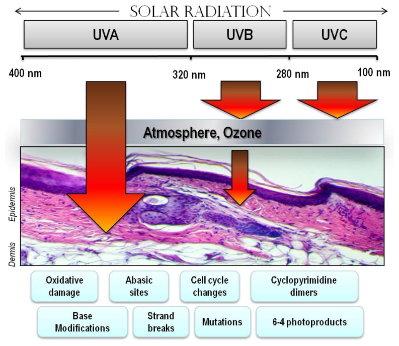
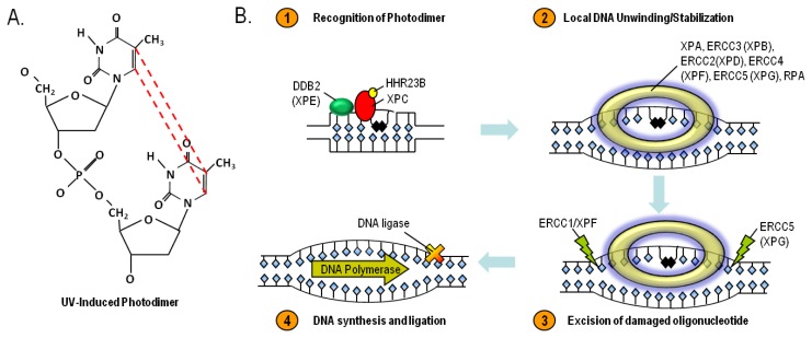
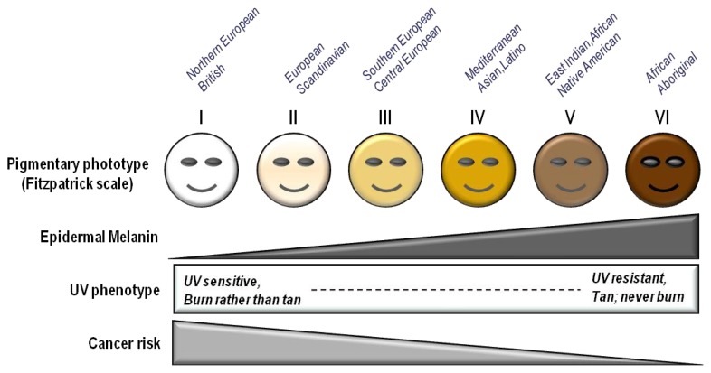
UV radiation (UV) is classified as a "complete carcinogen" because it is both a mutagen and a non-specific damaging agent and has properties of both a tumor initiator and a tumor promoter. In environmental abundance, UV is the most important modifiable risk factor for skin cancer and many other environmentally-influenced skin disorders. However, UV also benefits human health by mediating natural synthesis of vitamin D and endorphins in the skin, therefore UV has complex and mixed effects on human health. Nonetheless, excessive exposure to UV carries profound health risks, including atrophy, pigmentary changes, wrinkling and malignancy. UV is epidemiologically and molecularly linked to the three most common types of skin cancer, basal cell carcinoma, squamous cell carcinoma and malignant melanoma, which together affect more than a million Americans annually. Genetic factors also influence risk of UV-mediated skin disease. Polymorphisms of the melanocortin 1 receptor (MC1R) gene, in particular, correlate with fairness of skin, UV sensitivity, and enhanced cancer risk. We are interested in developing UV-protective approaches based on a detailed understanding of molecular events that occur after UV exposure, focusing particularly on epidermal melanization and the role of the MC1R in genome maintenance.


Figures

Figure 1
Figure 1
Epidermal structure and keratinocyte differentiation. The epidermis is a self-renewing tissue composed mainly of keratinocytes in various stages of terminal differentiation. Keratinocytes are produced in the stratum basale (basal layer), and move outward through the epidermis, undergoing a programmed series of differentiation involving enucleation and accumulation of cytokeratins and tight junctions with each other. Keratinocytes also receive melanin from melanocytes in the form of pre-packaged organelles termed melanosomes. The basic layers from the basement membrane outward are the stratum basale, stratum spinosum, stratum granulosum, and the stratum corneum, each identified by the morphology and differentiation state of the keratinocyte as indicated by expression of cytokeratins and other proteins.
Figure 2
Figure 2
Melanin Biosynthesis. Melanin, a large bioaggregate composed of pigmented chemical species, is found in two major forms: the brown/black highly UV-protective “eumelanin” pigment and the red/blonde UV-permeable “pheomelanin”. Both eumelanin and pheomelanin are derived from the amino acid tyrosine. Tyrosinase is the enzyme that catalyzes the rate-limiting synthetic reaction for both melanin species and when defective causes albinism. Incorporation of cysteine into pheomelanin results in the retention of sulfur into the pigment, which yields a light color to the final melanin product and may contribute to oxidative injury in the skin. The melanocyte stimulating hormone (MSH)–melanocortin 1 receptor (MC1R) signaling axis is a major determinant of the type and amount of melanin produced by melanocytes in the skin.
Figure 3
Figure 3
Electromagnetic spectrum of visible and UV radiation and biologic effects on the skin. Solar UV radiation can be subdivided into UVA, UVB and UVC components, however because of atmospheric ozone that absorbs UVC, ambient sunlight is predominantly UVA (90%–95%) and UVB (5%–10%). UV penetrates the skin in a wavelengthdependent manner. Longer wavelength UVA penetrates deeply into the dermis reaching well into the dermis. In contrast, UVB is almost completely absorbed by the epidermis, with comparatively little reaching the dermis. UVA is efficient at generating reactive oxygen species that can damage DNA via indirect photosensitizing reactions. UVB is directly absorbed by DNA which causes molecular rearrangements forming the specific photoproducts such as cyclobutane dimers and 6–4 photoproducts. Mutations and cancer can result from many of these modifications to DNA.
Figure 4
Figure 4
Mechanisms of the physiologic tanning response. Hormonal interactions between epidermal keratinocytes and melanocytes mediate much of the cutaneous melanization response. DNA and cellular damage in keratinocytes up-regulates transcription of the pro-opiomelanocortin (POMC) gene which encodes production and secretion of melanocyte stimulating hormone (α-MSH). α-MSH binding to melanocortin 1 receptor (MC1R) on melanocytes in the basal epidermis generates the second messenger cAMP via interactions between MC1R and adenylyl cyclase, and leads to activation of protein kinase A and the cAMP responsive binding element (CREB) and microphthalmia (Mitf) transcription factors. CREB and Mitf directly enhance melanin production by raising levels of tyrosinase and other melanin biosynthetic enzymes. Thus, MSH-MC1R signaling leads to enhanced pigment synthesis by melanocytes and accumulation of melanin by epidermal keratinocytes. By this mechanism, the skin is better protected against UV insults. Of note, UV-induced pigmentation may also occur through other signaling pathways as well as direct effects of UV on melanocytes, and there is some disagreement in the field over the role of epidermal MSH in the adaptive pigmentary response.
Figure 5
Figure 5
UV generates oxidative free radicals. UV photons interact with atomic oxygen to promote formation of free radical derivatives such as superoxide, hydrogen peroxide and the highly reactive hydroxyl radical. Free radicals avidly attack macromolecules such as protein, lipid, RNA and DNA, altering their structure and interfering with their function. Detoxifying and protective enzymes such as superoxide dismutase, catalase and glutathione peroxidase detoxify and reduce levels of oxidative species in the cell.
Figure 6
Figure 6
UV-induced cyclobutane dimers- structure (A) and repair by the Nucleotide Excision DNA Repair (NER) pathway (B). The NER pathway is mediated by at least eight enzymes that work together to identify bulky DNA lesions that distort the structure of the double helix, excise the damaged portion and replace the excised region by DNA synthesis directed by the complementary strand. Homozygous deficiency in any one of the NER enzymes leads to the clinical condition known as Xeroderma Pigmentosum (XP). Although not shown, NER can also be initiated in actively transcribed regions of the genome by involvement of the Cockayne syndrome proteins A and B.
Figure 7
Figure 7
Influence of pigmentation on skin cancer risk. Fair-skinned individuals with low levels of melanin in the epidermis display a UV sensitive phenotype, tending to burn rather than tan, after UV exposure. Recent data suggest that mutations that contribute to fair complexion and tanning impairment, specifically signaling defects in the melanocortin 1 receptor (MC1R), may also be associated with less efficient DNA repair in melanocytes. MC1R-defective individuals not only suffer higher realized doses of UV radiation because their skin is less able to block UV photons, but they may also accumulate more mutations from UV exposure because of defective DNA repair.

References

    1. Elwood J.M., Jopson J. Melanoma and sun exposure: An overview of published studies. Int. J. Cancer. 1997;73:198–203. - PubMed
    1. Lowe N.J. An overview of ultraviolet radiation, sunscreens, and photo-induced dermatoses. Dermatol. Clin. 2006;24:9–17. - PubMed
    1. Slominski A., Wortsman J. Neuroendocrinology of the skin. Endocr. Rev. 2000;21:457–487. - PubMed
    1. Slominski A., Wortsman J., Luger T., Paus R., Solomon S. Corticotropin releasing hormone and proopiomelanocortin involvement in the cutaneous response to stress. Physiol. Rev. 2000;80:979–1020. - PubMed
    1. Fuchs E. Scratching the surface of skin development. Nature. 2007;445:834–842. - PMC - PubMed

Publication types