. 2013 Aug 14;32(16):2204-16.
doi: 10.1038/emboj.2013.133. Epub 2013 Jun 7.

XIAP inhibits autophagy via XIAP-Mdm2-p53 signalling

Affiliations

XIAP inhibits autophagy via XIAP-Mdm2-p53 signalling

Xing Huang et al. EMBO J. .

Abstract

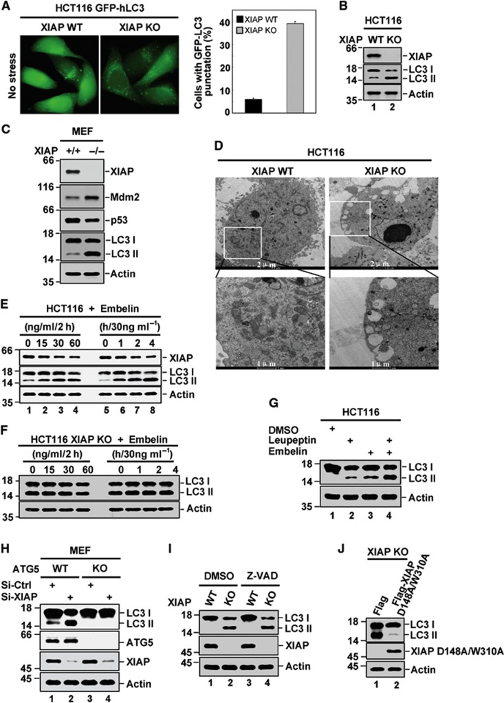
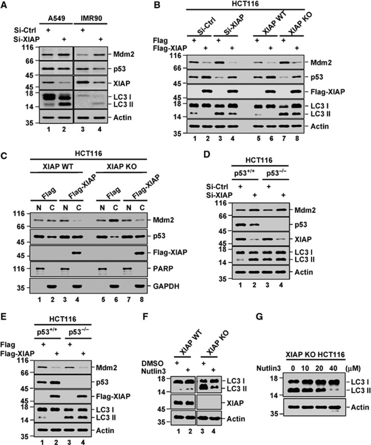
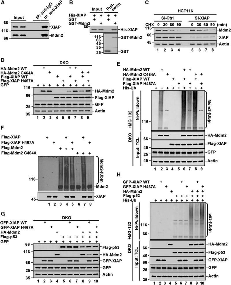
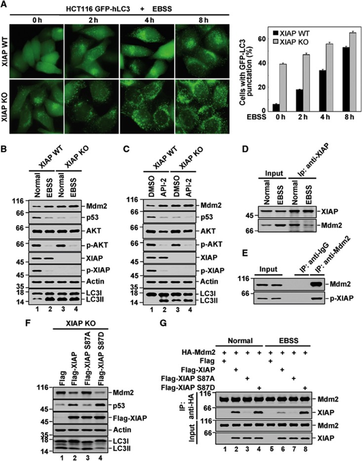
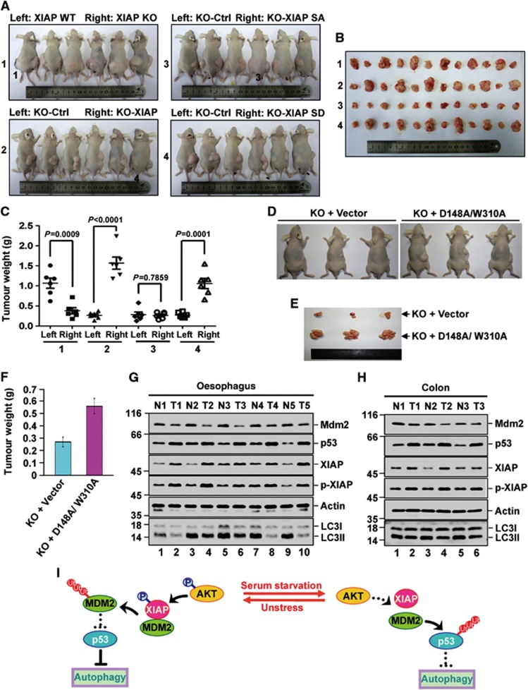
The primary role of autophagy is adaption to starvation. However, increasing evidence suggests that autophagy inhibition also plays an important role in tumorigenesis. Upregulation of X-linked inhibitor of apoptosis (XIAP) has been associated to a variety of human cancers, yet the underlying mechanisms remain obscure. Here, we report that XIAP suppresses autophagy by exerting a previously unidentified ubiquitin E3 ligase activity towards Mdm2, which is a negative regulator of p53. XIAP controls serum starvation-induced autophagy downstream of the PI3K/Akt pathway. In mouse models, inhibition of autophagy by XIAP promotes tumorigenecity of HCT116 cells. XIAP-mediated autophagy inhibition is also largely validated in clinical tumour samples. These findings reveal a novel XIAP-Mdm2-p53 pathway that mediates the inhibition of autophagy, by which XIAP may contribute to tumorigenesis.


Conflict of interest statement

The authors declare that they have no conflict of interest.

Figures

Figure 1
Figure 1
XIAP inhibits autophagy. (A) Representative images of GFP-LC3 staining in HCT116 XIAP WT and XIAP KO cells with stable expression of GFP-LC3. Quantification of LC3 punctate cells was shown on the right. The data are represented as means±s.d. of three independent experiments. (B) Endogenous LC3 expression in HCT116 XIAP WT and XIAP KO cells was analysed by western blotting with anti-LC3 antibody. The data are representative of three biological replicates. The ratio of LCII/LC3I to actin is presented in Supplementary Figure S1A. (C) Lysates from XIAP+/+ and XIAP−/− mouse embryonic fibroblasts were analysed by western blotting with the indicated antibodies. The data are representative of two biological replicates. (D) Autophagic vesicles in HCT116 XIAP WT and XIAP KO cells were evaluated by electron microscopy. The images are representative of three biological replicates. (E) HCT116 cells were treated with Embelin as indicated. LC3-II accumulation was determined by western blot analysis with anti-LC3 antibody. The data are representative of three biological replicates. The ratio of LCII/LC3I to actin is presented in Supplementary Figure S1B. (F) HCT116 XIAP KO cells were treated with Embelin as indicated. LC3-II accumulation was determined by western blot analysis with anti-LC3 antibody. The data are representative of three biological replicates. (G) HCT116 cells were treated with Leupeptin and Embelin in the indicated combinations. Cell lysates were analysed by western blotting. The data are representative of three biological replicates. The ratio of LCII/LC3I to actin is presented in Supplementary Figure S1D. (H) MEF ATG5 WT and ATG5 KO cells were individually transfected with XIAP-specific or control siRNAs. Forty-eight hours after transfection, cell lysates were analysed by western blotting with the indicated antibodies. The data are representative of three biological replicates. The ratio of LCII/LC3I to actin is presented in Supplementary Figure S1E. (I) HCT116 XIAP WT and XIAP KO cells were treated with 20 μM Z-VAD-FMK or DMSO for 2 h. Cell lysates were then analysed by western blotting. The data are representative of three biological replicates. The ratio of LCII/LC3I to actin is presented in Supplementary Figure S1F. (J) HCT116 XIAP KO cells were transfected with either Flag-XIAP (D148A/W310A) or control plasmid. Twenty-four hours after transfection, cell lysates were subjected to western blot analysis with the indicated antibodies. The data are representative of three biological replicates. The ratio of LCII/LC3I to actin is presented in Supplementary Figure S1G.
Figure 2
Figure 2
XIAP inhibits autophagy via the Mdm2-p53 pathway. (A) A549 and IMR90 cells were transfected with XIAP-specific or control siRNAs. Cell lysates were subjected to western blot analysis with the indicated antibodies. The data are representative of two biological replicates. The ratio of LCII/LC3I to actin is presented in Supplementary Figure S2A. (B) HCT116 cells expressing XIAP-specific or control siRNAs, HCT116 XIAP WT and XIAP KO cells were individually transfected with Flag-XIAP or control vector. Cell lysates were analysed by western blotting. The data are representative of three biological replicates. The ratio of LCII/LC3I to actin is presented in Supplementary Figure S2D. (C) HCT116 XIAP WT and XIAP KO cells were individually transfected with Flag-XIAP or control vector. Cytoplasmic/nuclear fractionation was performed to analyse cellular localization of Mdm2 and p53. PARP and GAPDH were used as nuclear (N) and cytoplasmic (C) fraction markers, respectively. The data are representative of three biological replicates. (D) HCT116 p53+/+ and p53−/− cells were individually transfected with XIAP-specific or control siRNAs. LC3 conversion was detected by western blot analysis. The data are representative of three biological replicates. The ratio of LCII/LC3I to actin is presented in Supplementary Figure S2E. (E) HCT116 p53+/+ and p53−/− cells were individually transfected with Flag-XIAP or control vector. LC3 conversion was detected by western blot analysis. The data are representative of three biological replicates. The ratio of LCII/LC3I to actin is presented in Supplementary Figure S2F. (F) HCT116 XIAP WT and XIAP KO cells treated with 10 μM Nutlin3 for 6 h. LC3 conversion was evaluated by western blot analysis. The data are representative of three biological replicates. The ratio of LCII/LC3I to actin is presented in Supplementary Figure S2H. (G) HCT116 XIAP KO cells were treated with the indicated amounts of Nutlin for 8 h. Cell lysates were analysed by western blotting with anti-LC3 antibody. The data are representative of three biological replicates.
Figure 3
Figure 3
XIAP is a novel E3 ligase for Mdm2. (A) Lysates from HCT116 cells were immunoprecipitated separately with anti-XIAP antibody and an isotype-matched control IgG. Immunoprecipitates and input were analysed by western blotting. The data are representative of two biological replicates. (B) Recombinant His-XIAP protein was incubated separately with purified GST and GST-Mdm2 fusion proteins on glutathione beads for 4 h followed by western blot analysis using anti-His antibody. GST and GST-Mdm2 were analysed by Coomassie blue staining. The data are representative of two biological replicates. (C) HCT116 cells were treated with XIAP-specific or control siRNAs. Twenty-four hours later, cells were cultured in the presence of 50 μg ml−1 cycloheximide for the indicated periods of time, and subsequently analysed by western blotting. We should mention that amounts of cell lysates were adjusted to achieve similar expression levels of Mdm2 at time 0, while the same amounts of cell lysates were used to examine levels of XIAP and actin. The data are representative of three biological replicates. The ratio of Mdm2 to actin is presented in Supplementary Figure S3B. (D) p53−/−Mdm2−/− MEF cells were co-transfected with the indicated Mdm2, XIAP, and p53 constructs. Cell lysates were analysed by western blotting with the indicated antibodies. We should mention that XIAP H467A expressing plasmid was always used less than XIAP expressing plasmid to ensure expression levels of XIAP and XIAP H467A were similar. The data are representative of three biological replicates. (E) p53−/−Mdm2−/− MEF cells were transfected with the indicated plasmids. Twenty-four hours after transfection, cells were treated with 20 μM MG-132 for additional 4 h. Cell lysates were denatured before proteins conjugated to His-ubiquitin were pulled down by Ni2+-NTA beads. The bead-bound proteins and total cell lysates (TCLs) were analysed by western blot with anti-Mdm2 antibody. The data are representative of three biological replicates. (F) In all, 2 μM Mdm2 or its C464A mutant and 1 μM XIAP or its H467A mutant proteins were incubated with 100 nM E1, 2 μM E2 and 200 μM Ub in a total 20 μl in vitro ubiquitination reaction buffer at 37°C for 1 h. The reaction mixtures were analysed by western blotting with anti-Mdm2 antibody. The data are representative of three biological replicates. (G) p53−/−Mdm2−/− MEF cells were transfected with the indicated plasmids. Cell lysates were analysed by western blotting. The data are representative of three biological replicates. (H) p53−/−Mdm2−/− MEF cells were transfected with the indicated plasmids. Twenty-four hours after transfection, cells were treated with 20 μM MG-132 for additional 4 h. Cell lysates were denatured before proteins conjugated to His-ubiquitin were pulled down by Ni2+-NTA beads. The bead-bound proteins were analysed by western blotting with anti-p53 antibody. The data are representative of three biological replicates.
Figure 4
Figure 4
XIAP regulates serum starvation-induced autophagy. (A) HCT116 XIAP WT and XIAP KO cells were treated with EBSS for the indicated periods of time. GFP-LC3 puncta formation was observed by a microscope. Quantification of LC3 punctate cells was shown on the right. The data are represented as means±s.d. of three independent experiments. (B) HCT116 XIAP WT and XIAP KO cells were treated with EBSS for 4 h followed by western blot analysis with the indicated antibodies. The data are representative of three biological replicates. The ratio of LCII/LC3I to actin is presented in Supplementary Figure S6B. (C) HCT116 XIAP WT and XIAP KO cells were treated with 1 μM API-2 for 6 h. Cell lysates were analysed by western blotting. The data are representative of three biological replicates. The ratio of LCII/LC3I to actin is presented in Supplementary Figure S6C. (D) HCT116 cells were treated with or without EBSS for 4 h. Cell lysates were subjected to immunoprecipitation with anti-XIAP antibody followed by western blot analysis. The data are representative of two biological replicates. (E) Lysates from HCT116 cells were immunoprecipitated with anti-Mdm2 antibody. Immunoprecipitates and cell lysates were analysed by western blotting with anti-p-XIAP antibody. The data are representative of two biological replicates. (F) HCT116 XIAP KO cells were transfected with Flag-XIAP, Flag-XIAP S87A, Flag-XIAP S87D or control vector. Twenty-four hours after transfection, cells were harvested. LC3 conversion was evaluated by western blot analysis. The data are representative of three biological replicates. The ratio of LCII/LC3I to actin is presented in Supplementary Figure S6D. (G) p53−/−Mdm2−/− MEF cells were transfected with HA-Mdm2 plus Flag-XIAP, Flag-XIAP S87A or Flag-XIAP S87D. Twenty-four hours later, cells were treated with 20 μM MG-132 for another 4 h. Cell lysates were subjected to immunoprecipitation with anti-HA antibody. Immunoprecipitates were analysed by western blotting. The data are representative of two biological replicates.
Figure 5
Figure 5
XIAP-modulated autophagy is associated with the tumorigenecity. (AC) 1 × 10⁶ HCT116 XIAP WT and 1 × 10⁶ XIAP KO cells (group 1), 1 × 10⁶ HCT116 XIAP KO cells (KO-Ctrl) and 1 × 10⁶ HCT116 XIAP KO cells stably expressing wild-type XIAP (KO-XIAP) (group 2), 1 × 10⁶ HCT116 XIAP KO cells (KO-Ctrl) and 1 × 10⁶ HCT116 XIAP KO cells stably expressing XIAP S87A mutant (KO-XIAP SA) (group 3), or 1 × 10⁶ HCT116 XIAP KO cells (KO-Ctrl) and 1 × 10⁶ HCT116 XIAP KO cells stably expressing XIAP S87D mutant (KO-XIAP SD) (group 4) were individually injected to the left flank (Left) and right flank (Right) of nude mice as indicated. Five weeks after injection, the mice were sacrificed and photographed (A). The volumes of all flank tumours excised from six mice in each group were compared (n=6) (B). Tumour weights were represented as means±s.d. from six mice in each group (C). (DF) 1 × 10⁶ HCT116 XIAP KO cells or 1 × 10⁶ HCT116 XIAP KO cells stably expressing XIAP D148A/310A were injected to the right flank of nude mice as indicated (n=3). Five weeks after injection, the mice were sacrificed and photographed (D). The volumes of excised tumours were shown (E). Tumour weights were represented as means±s.d. from three mice in each group (F). (G, H) The human tumour tissues (T) from Oesophagus (G) and Colon (H) and their adjacent normal tissues (N) were homogenized for protein extraction. Protein extracts were analysed by western blotting with the indicated antibodies. The data are representative of three biological replicates. The blots in (G, H) were qualified and the ratio of LC3II/LC3I to actin was then calculated and shown in Supplementary Figure 9A and B. (I) The schematic model of autophagic regulation by XIAP is shown. Under non-stressed condition, phosphorylated XIAP interacts with Mdm2 and mediates its rapid degradation, thus maintaining the relative high levels of p53 and inhibiting basal levels of autophagy. In response to serum starvation, XIAP undergoes dephosphorylation due to AKT inhibition, thereby promoting the dissociation of XIAP from Mdm2. This in turn accelerates the degradation of p53 and facilitates serum starvation-induced autophagy.

Comment in

  • XIAP: inhibitor of two worlds.
    Merlo P, Cecconi F. Merlo P, et al. EMBO J. 2013 Aug 14;32(16):2187-8. doi: 10.1038/emboj.2013.152. Epub 2013 Jun 21. EMBO J. 2013. PMID: 23792426 Free PMC article.

Similar articles

Cited by

References

    1. Aita VM, Liang XH, Murty VV, Pincus DL, Yu W, Cayanis E, Kalachikov S, Gilliam TC, Levine B (1999) Cloning and genomic organization of beclin 1, a candidate tumor suppressor gene on chromosome 17q21. Genomics 59: 59–65 - PubMed
    1. Birnbaum MJ, Clem RJ, Miller LK (1994) An apoptosis-inhibiting gene from a nuclear polyhedrosis virus encoding a polypeptide with Cys/His sequence motifs. J Virol 68: 2521–2528 - PMC - PubMed
    1. Crighton D, Wilkinson S, O'Prey J, Syed N, Smith P, Harrison PR, Gasco M, Garrone O, Crook T, Ryan KM (2006) DRAM, a p53-induced modulator of autophagy, is critical for apoptosis. Cell 126: 121–134 - PubMed
    1. Crook NE, Clem RJ, Miller LK (1993) An apoptosis-inhibiting baculovirus gene with a zinc finger-like motif. J Virol 67: 2168–2174 - PMC - PubMed
    1. Damgaard RB, Nachbur U, Yabal M, Wong WW, Fiil BK, Kastirr M, Rieser E, Rickard JA, Bankovacki A, Peschel C, Ruland J, Bekker-Jensen S, Mailand N, Kaufmann T, Strasser A, Walczak H, Silke J, Jost PJ, Gyrd-Hansen M (2012) The ubiquitin ligase XIAP recruits LUBAC for NOD2 signaling in inflammation and innate immunity. Mol Cell 46: 746–758 - PubMed

Publication types

MeSH terms

Substances