. 2013 Dec;9(4):573-83.
doi: 10.1007/s11302-013-9368-1. Epub 2013 Jun 8.

Adenosine A2A receptor (A2AR) is a fine-tune regulator of the collagen1:collagen3 balance

Affiliations

Adenosine A2A receptor (A2AR) is a fine-tune regulator of the collagen1:collagen3 balance

Miguel Perez-Aso et al. Purinergic Signal. 2013 Dec.

Abstract

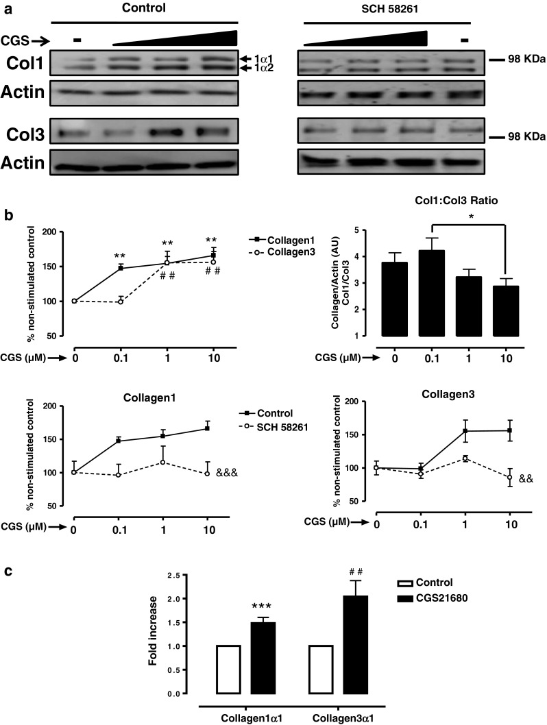
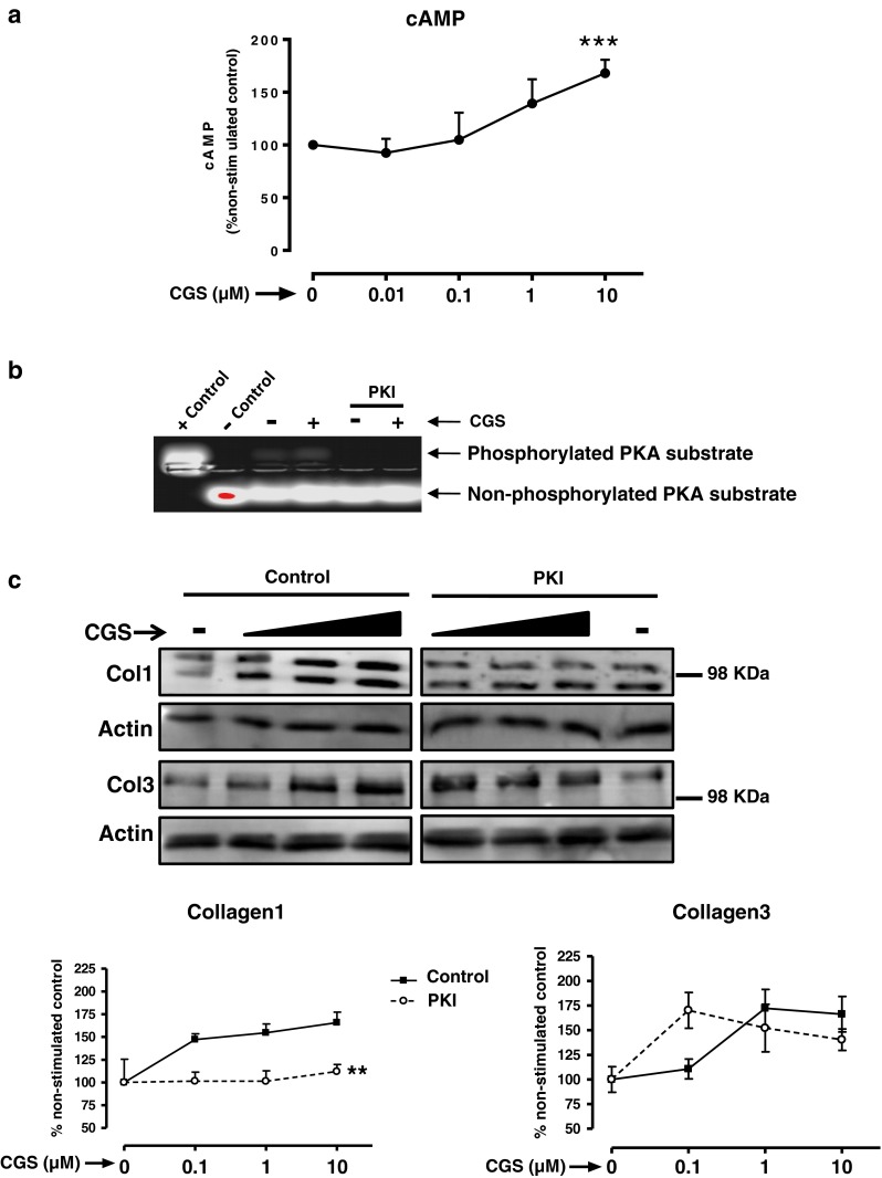
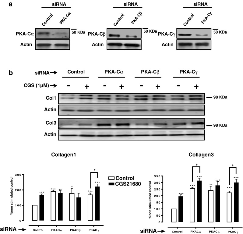
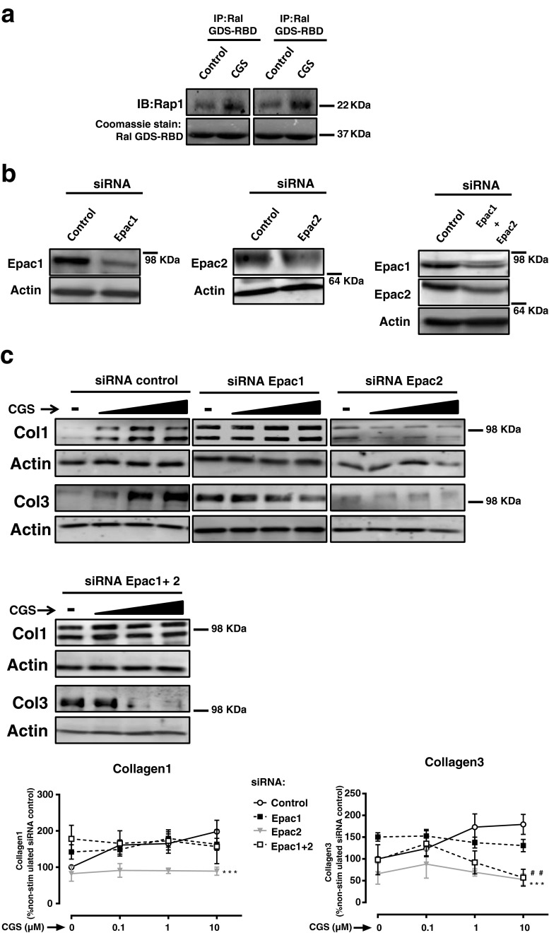
Adenosine is a potent endogenous anti-inflammatory and immunosuppressive metabolite that is a potent modulator of tissue repair. However, the adenosine A2A receptor (A2AR)-mediated promotion of collagen synthesis is detrimental in settings such as scarring and scleroderma. The signaling cascade from A2AR stimulation to increased collagen production is complex and obscure, not least because cAMP and its downstream molecules PKA and Epac1 have been reported to inhibit collagen production. We therefore examined A2AR-stimulated signaling for collagen production by normal human dermal fibroblasts (NHDF). Collagen1 (Col1) and collagen3 (Col3) content after A2AR activation by CGS21680 was studied by western blotting. Contribution of PKA and Epac was analyzed by the PKA inhibitor PKI and by knockdowns of the PKA-Cα, -Cβ, -Cγ, Epac1, and Epac2. CGS21680 stimulates Col1 expression at significantly lower concentrations than those required to stimulate Col3 expression. A2AR stimulates Col1 expression by a PKA-dependent mechanism since PKA inhibition or PKA-Cα and -Cβ knockdown prevents A2AR-mediated Col1 increase. In contrast, A2AR represses Col3 via PKA but stimulates both Col1 and Col3 via an Epac2-dependent mechanism. A2AR stimulation with CGS21680 at 0.1 μM increased Col3 expression only upon PKA blockade. A2AR activation downstream signaling for Col1 and Col3 expression proceeds via two distinct pathways with varying sensitivity to cAMP activation; more highly cAMP-sensitive PKA activation stimulates Col1 expression, and less cAMP-sensitive Epac activation promotes both Col1 and Col3 expression. These observations may explain the dramatic change in Col1:Col3 ratio in hypertrophic and immature scars, where adenosine is present in higher concentrations than in normal skin.


Figures

Fig. 1
Fig. 1
A2AR activation increase collagen I and III with different potency. a NHDF cells were incubated with increasing concentrations of the A2AR agonist CGS21680 during 24 h, with or without pre-incubation with the A2AR selective antagonist SCH58261 0.1 μM. Black triangle represents the increasing concentrations of CGS21680 0.1–1–10 μM. b Densitometry of bands showing percent change and Col1:Col3 ratio. Data represent means ± SEM of more than ten independent experiments, and statistics was performed by ANOVA followed by Newman–Keuls post-test, Col1 **P < 0.01 and Col3 ## P < 0.01 vs. non-stimulated control; *P < 0.05 CGS21680 0.1 vs. 10 μM; or by two-way ANOVA, &&& P < 0.001 and && P < 0.01 SCH58261 vs. control. c mRNA for collagen1α1 and collagen3α1 was measured by real-time RT-PCR after a 24-h stimulation with CGS21680 1 μM. Data represent means ± SEM of four independent experiments, and statistical analysis was performed by Student's t test, collagen1α1 ***P < 0.001 and collagen3α1 ## P < 0.01 vs. non-stimulated control
Fig. 2
Fig. 2
PKA activates collagen I but inhibits collagen III. a Intracellular cAMP levels were measured as described in “Methods” section. Statistical analysis was performed by Student's t test, ***P < 0.001 vs. non-stimulated. b PKA activity measurement; when indicated, 1 h prior to CGS21680 stimulation, NHDF were pre-incubated with the PKA inhibitor PKI (10 μg/ml), and PKA activity was analyzed as described in “Methods” section. c Cells were pre-incubated with the PKA inhibitor PKI (10 μg/ml) before CGS21680 addition and Col1 and Col3 expression analysis. Black triangle represents the increasing concentrations of CGS21680 0.1–1–10 μM. Statistical analysis was performed by two-way ANOVA, **P < 0.01 PKI vs. control. Data represent means ± SEM of three or more independent experiments
Fig. 3
Fig. 3
Impact of knockdown of the PKA catalytic subunits on Col1 and Col3 expression. a siRNAs for PKA-Cα, PKA-Cβ, and PKA-Cγ selectively reduce the protein expression when compared to control siRNA. b Impact of PKA-Cα, PKA-Cβ, and PKA-Cγ silencing on basal and CGS21680 1-μM-increased Col1 and Col3 expression. Statistics was performed by Student's t test, *P < 0.05, **P < 0.01, and ***P < 0.001 vs. non-stimulated control siRNA and # P < 0.01 CGS216280 vs. non-stimulated of the same siRNA. Data represent means ± SEM of three or more independent experiments
Fig. 4
Fig. 4
Impact of Epac1/2 knockdown on Col1 and Col3 expression. a Epac activity was analyzed after incubation with CGS21680 1 μM for 15 min. Immunoprecipitation with Ral GSD-RBD was performed followed by immunoblot with anti-Rap1, and Coomassie stain identified Ral GSD-RBD at the predicted molecular weight of 37 kDa. b siRNAs for Epac1, Epac2, and double knockdown Epac1 + 2 selectively reduce the protein expression when compared to control siRNA. c Impact of Epac1, Epac2 silencing, or double knockdown Epac1 + 2 on basal and CGS21680-increased Col1 and Col3 expression. Statistics was performed by the two-way ANOVA: Epac2 siRNA ***P < 0.001 and Epac1 + 2 siRNA ## P < 0.01 vs. control siRNA. Black triangle represents the increasing concentrations of CGS21680 0.1–1–10 μM, and data represent means ± SEM of three or more independent experiments

References

    1. Williams JC. Adenine nucleotide degradation by the obligate intracellular bacterium Rickettsia typhi. Infect Immun. 1980;28(1):74–81. - PMC - PubMed
    1. Hasko G, Cronstein BN. Adenosine: an endogenous regulator of innate immunity. Trends Immunol. 2004;25(1):33–39. doi: 10.1016/j.it.2003.11.003. - DOI - PubMed
    1. Ohta A, Sitkovsky M. Role of G-protein-coupled adenosine receptors in downregulation of inflammation and protection from tissue damage. Nature. 2001;414(6866):916–920. doi: 10.1038/414916a. - DOI - PubMed
    1. Stadelmann WK, Digenis AG, Tobin GR. Physiology and healing dynamics of chronic cutaneous wounds. Am J Surg. 1998;176(2A Suppl):26S–38S. doi: 10.1016/S0002-9610(98)00183-4. - DOI - PubMed
    1. Victor-Vega C, Desai A, Montesinos MC, Cronstein BN. Adenosine A2A receptor agonists promote more rapid wound healing than recombinant human platelet-derived growth factor (Becaplermin gel) Inflammation. 2002;26(1):19–24. doi: 10.1023/A:1014417728325. - DOI - PubMed

Publication types

MeSH terms

LinkOut - more resources