. 2013 Aug 1;73(15):4791-800.
doi: 10.1158/0008-5472.CAN-13-0587. Epub 2013 Jun 7.

Enhanced radiation sensitivity in HPV-positive head and neck cancer

Affiliations

Enhanced radiation sensitivity in HPV-positive head and neck cancer

Randall J Kimple et al. Cancer Res. .

Abstract

Patients with human papillomavirus (HPV+)-associated head and neck cancer (HNC) show significantly improved survival outcome compared with those with HPV-negative (HPV-) tumors. Published data examining this difference offers conflicting results to date. We systematically investigated the radiation sensitivity of all available validated HPV+ HNC cell lines and a series of HPV- HNC cell lines using in vitro and in vivo techniques. HPV+ HNCs exhibited greater intrinsic radiation sensitivity (average SF2 HPV-: 0.59 vs. HPV+: 0.22; P < 0.0001), corresponding with a prolonged G2-M cell-cycle arrest and increased apoptosis following radiation exposure (percent change 0% vs. 85%; P = 0.002). A genome-wide microarray was used to compare gene expression 24 hours following radiation between HPV+ and HPV- cell lines. Multiple genes in TP53 pathway were upregulated in HPV+ cells (Z score 4.90), including a 4.6-fold increase in TP53 (P < 0.0001). Using immortalized human tonsillar epithelial (HTE) cells, increased radiation sensitivity was seen in cell expressing HPV-16 E6 despite the effect of E6 to degrade p53. This suggested that low levels of normally functioning p53 in HPV+ HNC cells could be activated by radiation, leading to cell death. Consistent with this, more complete knockdown of TP53 by siRNA resulted in radiation resistance. These results provide clear evidence, and a supporting mechanism, for increased radiation sensitivity in HPV+ HNC relative to HPV- HNC. This issue is under active investigation in a series of clinical trials attempting to de-escalate radiation (and chemotherapy) in selected patients with HPV+ HNC in light of their favorable overall survival outcome.


Figures

Figure 1
Figure 1
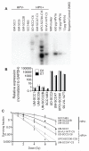
A) HPV-16 specific Southern blot of cell lines used confirmed the presence of HPV-16 DNA in UD-SCC2, UM-SCC47, UPCI-SCC90, and 93-VU-147T, but not UM-SCC1, UM-SCC6, UM-SCC22B, or SCC1483. C# refers to the clone number of each individual clonal population utilized. B) Quantitative RT-PCR of total cellular RNA for HPV-16 E6 and E7 confirmed the presence of RNA encoding HPV-16 E6 and HPV-16 E7 in HPV+, but not HPV− cells. Data shown are mean expression +/− SEM (each sample normalized to GAPDH) from three biologic replicates each run as three technical replicates. C. Increased radiation sensitivity in HPV-positive head and neck cancer cells. Clonogenic survival over a range of radiation doses for HPV+ cells (dashed lines, triangles) and HPV− (solid lines, circles) demonstrate significantly increased radiation sensitivity in HPV+ cells (n=6 per condition). Mean surviving fraction after 2 Gy is 22% for HPV+ vs. 59% for HPV− cells (p<0.0001).
Figure 2
Figure 2
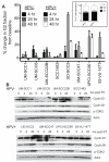
Radiation induced G2/M cell cycle arrest in HPV+ HNC. A) Absolute change in percentage of cells in G2/M at indicated time points after radiation (4 Gy) as assessed by propidium iodide staining and flow cytometry assessment. The baseline fraction of cells in G2/M is subtracted from indicated mean (n=3) population distribution. Inset: No difference in baseline cell cycle distribution between HPV+ and HPV− cells is observed. B) Immunoblot analysis showing activation of Cyclin B1 and phospho-CDK2, but not Cyclin E1 in HPV+ HNC cells following radiation (4 Gy).
Figure 3
Figure 3
Apoptosis in HPV+ HNC after radiation. A) Significantly increased caspase activity compared to baseline, unirradiated cells, was seen in HPV+, but not HPV− HNC. Shown is the mean percentage increase in caspase activity over baseline as measured by luminescent caspase activation assay at indicated times following a single 4 Gy dose of radiation (n=4-6 per condition). B) Annexin V and propidium iodide staining to identify an apoptotic population of cells demonstrated a significant increase in the proportion of cells in apoptosis (early + late) following a single 4 Gy dose of radiation in HPV+ HNC cells (n=3, 2-way ANOVA, p<0.0001).
Figure 4
Figure 4
HPV16 E6 modulation of radiation response. A) Increased TP53 RNA following radiation in HPV+ HNC cells. Data presents TP53 expression at indicated time points, normalized to unirradiated control cells for each individual cell line. B) Increased phospho- and total-p53 protein is seen in HPV+ HNC following radiation. Low levels of p53 in several cell lines necessitated prolonged exposures to identify protein bands. 25 μg of total protein was loaded into each well. C) The clonogenic capacity of human tonsillar epithelial (HTE) cells stably expressing vector alone (black squares, HPV-16 E6 (grey triangles) or HPV-16 E6/E7 (grey diamonds) was assessed by colony formation assay over a range of radiation doses showing that expression of HPV-16 E6 resulted in increased sensitivity to radiation. D) Increase in G2/M fraction in HPV16 E6 and E6/E7 expressing cells relative to baseline, unirradiated cells. Absolute change in percentage of cells in G2/M at indicated time points after radiation (4 Gy) as assessed by propidium iodide staining and flow cytometry assessment. The baseline fraction of cells in G2/M is subtracted from indicated mean (n=3) population distribution. E) Significantly increased caspase activity as measured by substrate cleavage compared to baseline, unirradiated cells, was seen in HPV16 E6 and E6/E7 expressing cells. F) 24 h after radiation, increased p21 RNA was detected by qRT-PCR in HPV+, but not HPV−, cells.
Figure 5
Figure 5
Critical role for p53 in radiation response of HPV+ HNC. A) Expression of HPV16 E6 results in decreased detectable p53 protein while use of p53 specific siRNA, but not scrambled or vehicle control, results in further loss of p53 expression in HTE HPV16 E6 expressing cells (left, 25 μg of total protein), UM-SCC47 cells (center, 75 μg of total protein) and 93-VU-147T cells (right, 25 μg of total protein). E) Knockdown of TP53 causes greater colony formation HTE +16E6, UM-SCC47, and 93-VU-147T cells (n=6 per condition). Compared to unirradiated cells the surviving fraction of scrambled siRNA pretreated cells was significantly lower than that of TP53 specific siRNA treated cells (*p<0.0001).
Figure 6
Figure 6
Increased radiation sensitivity observed in vivo using xenograft model system. Mice (n=10/condition) implanted with indicated cell lines (1×10⁶/site) in bilateral flanks were treated with four fractions of radiation (2 Gy) delivered over 2 weeks. Tumor volumes were measured, mean tumor volume +/− SEM is shown and curves were fit to an exponential growth equation and compared by the extra sum-of-squares f-test. Time to tumor quadrupling was calculated from the first day of treatment and graphed according to the Kaplan-Meier method. Curve comparisons for control vs. radiation treated mice were performed with the log-rank (Mantel Cox) test. A) HPV+ cell lines showed growth delay following radiation in 3 of 4 cell lines. B) No significant difference in time to tumor quadrupling was seen between control and radiation treated HPV− xenografts using the indicated treatment schedule (p=0.14). C) Radiation resulted in a significant delay in tumor quadrupling for HPV+ cells (p=0.0003).
Figure 7
Figure 7
Proposed model underlying increased sensitivity to radiation in HPV+ HNC. In HPV− HNC, mutations in either TP53, or alteration in p53 signaling bypass the cell cycle arrest typically induced following radiation. While HPV E6 in HPV+ HNC degrades p53, low levels of wild-type p53 remain and can be activated by radiation-induced DNA damage resulting in partial arrest and increased cell death.

References

    1. Gillison ML, Koch WM, Capone RB, Spafford M, Westra WH, Wu L, et al. Evidence for a causal association between human papillomavirus and a subset of head and neck cancers. J Natl Cancer Inst. 2000;92:709–20. - PubMed
    1. Chaturvedi AK, Engels EA, Anderson WF, Gillison ML. Incidence Trends for Human Papillomavirus-Related and -Unrelated Oral Squamous Cell Carcinomas in the United States. J Clin Oncol. 2008;26:612–9. - PubMed
    1. Rischin D, Young RJ, Fisher R, Fox SB, Le QT, Peters LJ, et al. Prognostic significance of p16INK4A and human papillomavirus in patients with oropharyngeal cancer treated on TROG 02.02 phase III trial. J Clin Oncol. 2010;28:4142–8. - PMC - PubMed
    1. Fakhry C, Westra WH, Li S, Cmelak A, Ridge JA, Pinto H, et al. Improved Survival of Patients With Human Papillomavirus-Positive Head and Neck Squamous Cell Carcinoma in a Prospective Clinical Trial. J Natl Cancer Inst. 2008;100:261–9. - PubMed
    1. Lindel K, Beer KT, Laissue J, Greiner RH, Aebersold DM. Human papillomavirus positive squamous cell carcinoma of the oropharynx: a radiosensitive subgroup of head and neck carcinoma. Cancer. 2001;92:805–13. - PubMed

Publication types

Substances