. 2013 Aug 1;73(15):4781-90.
doi: 10.1158/0008-5472.CAN-13-0566. Epub 2013 Jun 7.

PanIN-specific regulation of Wnt signaling by HIF2α during early pancreatic tumorigenesis

Affiliations

PanIN-specific regulation of Wnt signaling by HIF2α during early pancreatic tumorigenesis

Angela Criscimanna et al. Cancer Res. .

Abstract

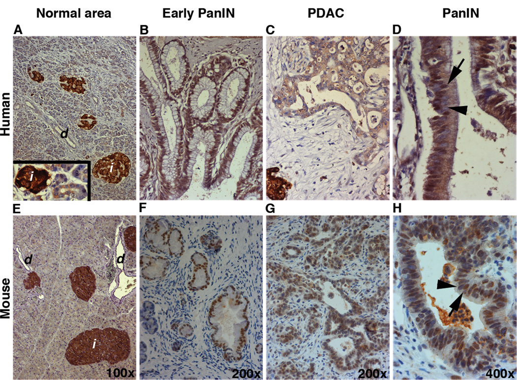
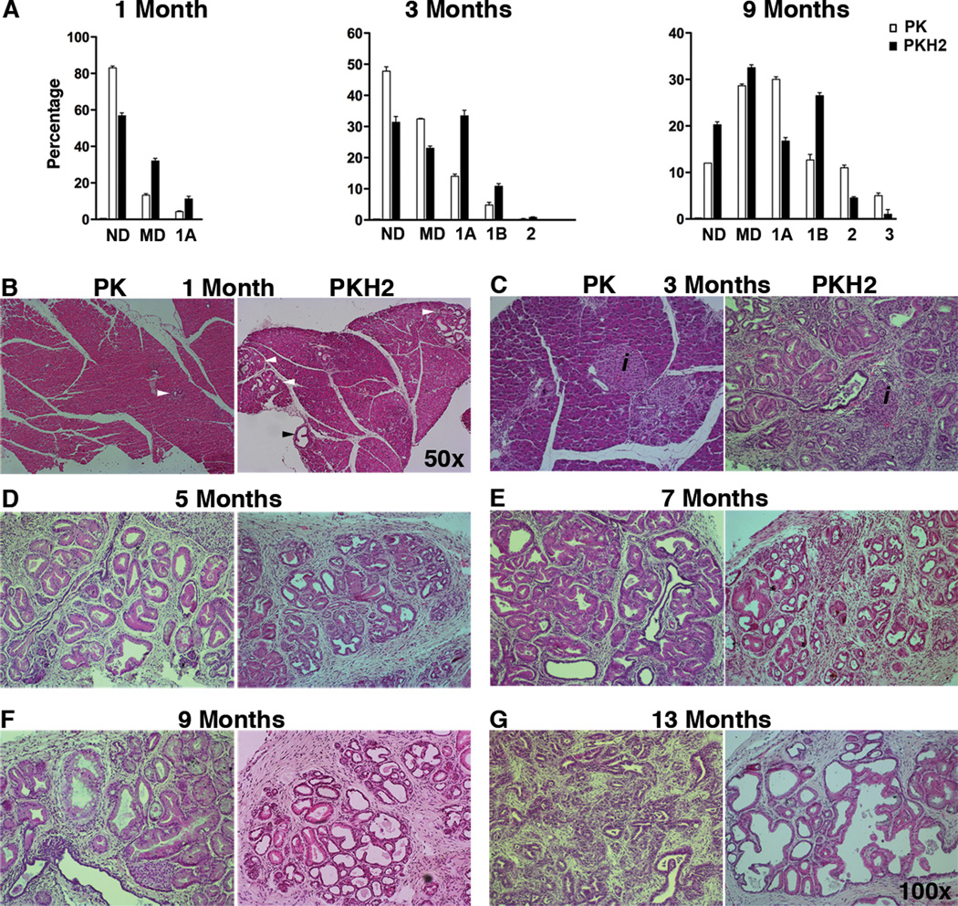
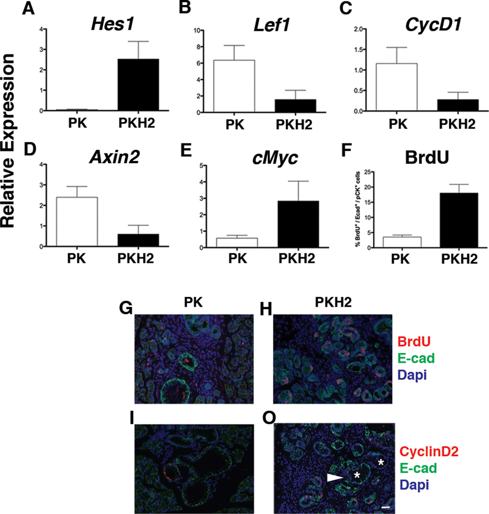
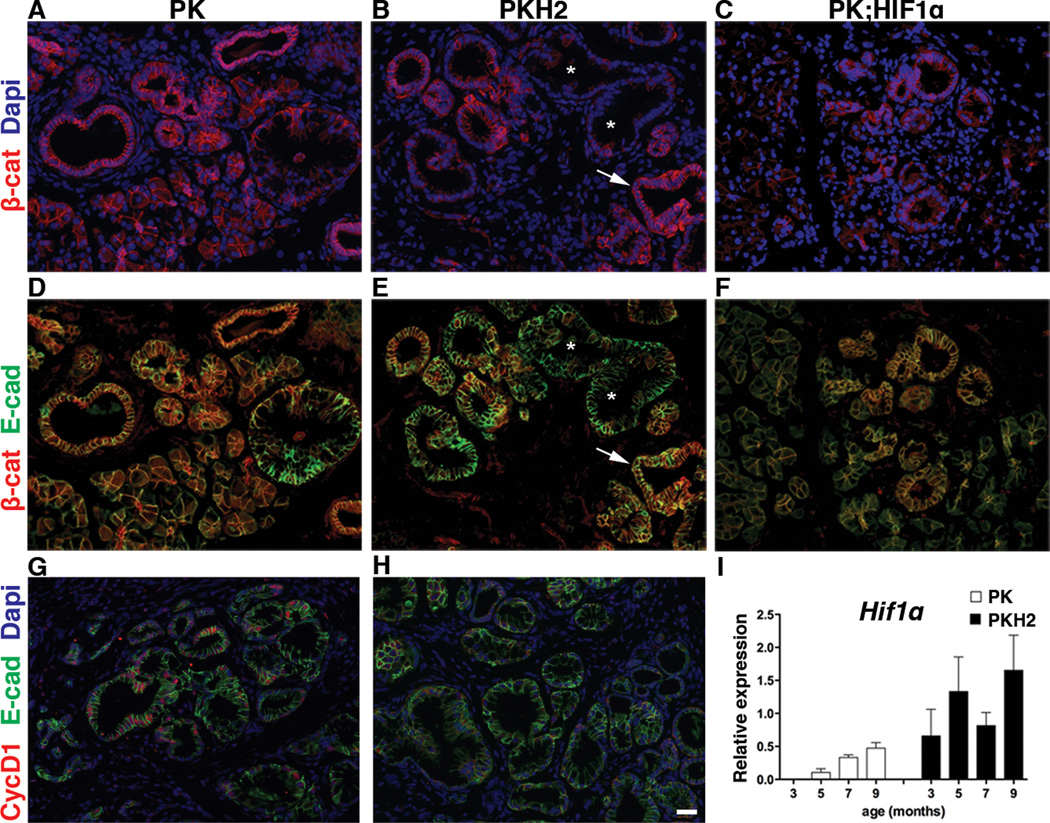
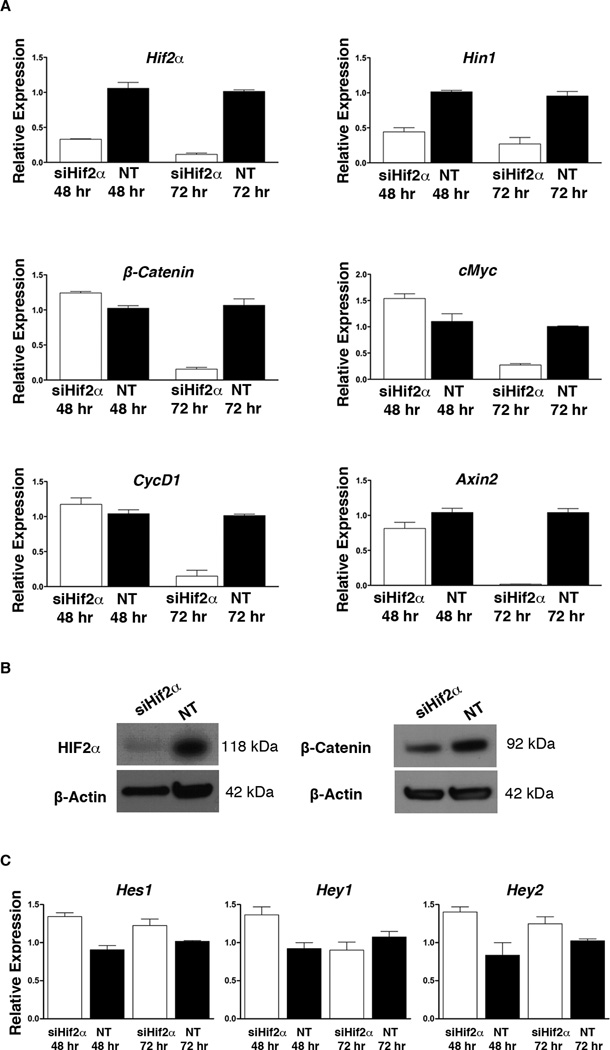
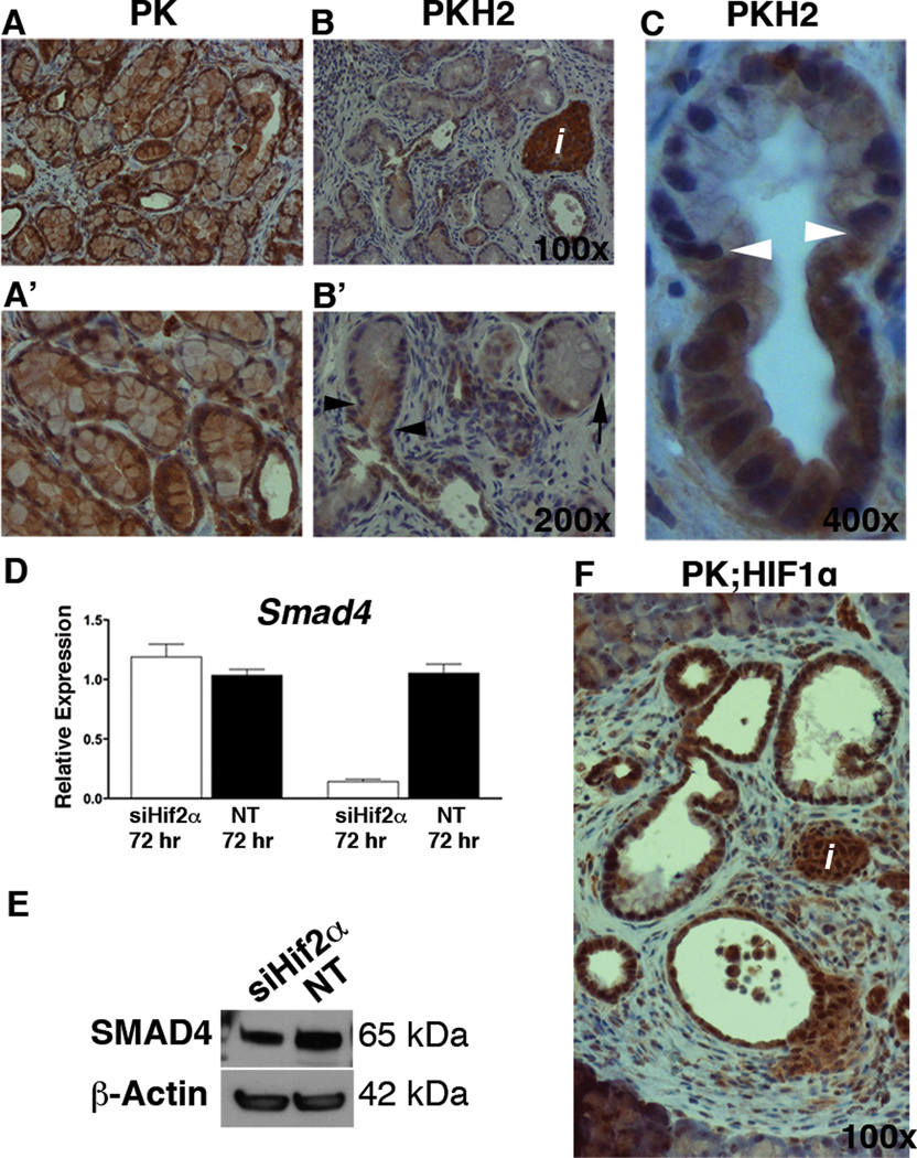
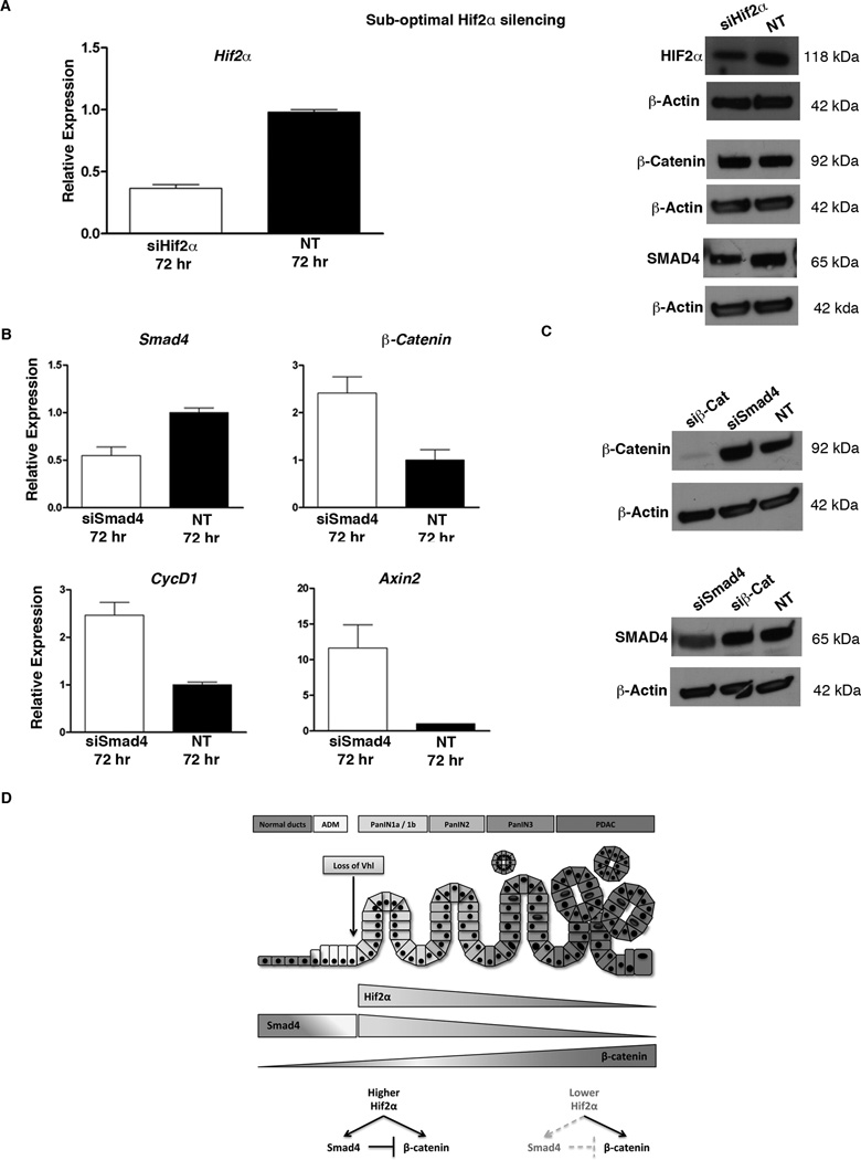
Hypoxia promotes angiogenesis, proliferation, invasion, and metastasis of pancreatic cancer. Essentially, all studies of the hypoxia pathway in pancreatic cancer research to date have focused on fully malignant tumors or cancer cell lines, but the potential role of hypoxia inducible factors (HIF) in the progression of premalignant lesions has not been critically examined. Here, we show that HIF2α is expressed early in pancreatic lesions both in human and in a mouse model of pancreatic cancer. HIF2α is a potent oncogenic stimulus, but its role in Kras-induced pancreatic neoplasia has not been discerned. We used the Ptf1aCre transgene to activate Kras(G12D) and delete Hif2α solely within the pancreas. Surprisingly, loss of Hif2α in this model led to markedly higher, rather than reduced, number of low-grade pancreatic intraepithelial neoplasia (mPanIN) lesions. These lesions, however, failed to progress to high-grade mPanINs, and displayed exclusive loss of β-catenin and SMAD4. The relationship among HIF2α, β-catenin, and Smad4 was further confirmed in vitro, where silencing of Hif2α resulted in reduced β-catenin and Smad4 transcript levels. Thus, with oncogenic Ras expressed in the pancreas, HIF2α modulates Wnt-signaling during mPanIN progression by maintaining appropriate levels of both Smad4 and β-catenin.


Conflict of interest statement

The authors disclose no conflicts.

Figures

Figure 1
Figure 1
HIF2α expression is gradually decreased during malignant progression. Immunohistochemical analyses for detection of HIF2α in human A–D and PK (E–H) pancreas. In the histologically normal pancreatic tissues in both human (A) and mice (E) HIF2α was absent in the acinar and ductal compartments. Inset in (A) shows acinar-to-ductal metaplastic structure with HIF2α-negative acinar cells turning into HIF2α-positive metaplastic ducts (arrowhead). HIF2α could be detected in early PanINs (B, F), whereas it’s expression was significantly reduced in more advanced lesions (C, G). Both human (D) and mouse (H) PanINs are composed of heteregenous populations of HIF2α-positive (arrows in G and H) and HIF2α-negative (arrowheads in G and H) cells. d: duct; i: islet.
Figure 2
Figure 2
HIF2α is required for mPanIN progression. (A) Higher early mPanIN incidence was observed in PKH2 pancreas. Columns, percentages (mean ± SE) of normal ducts, metaplastic ducts, and mPanINs by grade per total ductal structures in our two genotypes at 1, 3, and 9 months of age (n=5 for each cohort). (B–G) Representative tissues from PK and PKH2 collected at 1 (B), 3 (C), 5 (D), 7 (E), 9 (F) and 13 (G) months of age were stained for H&E. Arrowheads show the lesions on the sections. ND: Normal Ducts; MD: Metaplastic Ducts; 1A, 1B, 2, 3: mPanINs1A-3.
Figure 3
Figure 3
Decreased Wnt signaling in PKH2 pancreas. qRT-PCR analysis of RNA extracted from 3 months old PK and PKH2 whole pancreas shows higher Notch activity as evident by upregulation of Hes1 (A) and down regulation of Wnt-target genes in PKH2 pancreas (n=5 for each cohort). Bars represent gene expression (mean ± SE) relative to GAPDH. (B–E) Highly proliferative metaplastic PKH2 ducts. (F–J) Quantification of the proliferation rate showed a four-fold increase in BrdU incorporation in the PKH2 metaplastic ducts (F). Immunofluorescent analyses of 3 month old PK (G, I) and PKH2 (H, J) pancreas using antibodies against E-cadherin and BrdU (G, H) or E-cadherin and CyclinD2 (I, J) showed few CyclinD2+ or BrdU+ cells within PKH2 ducts (arrowhead) but not in mPanINs (Asterisks). Scale bar 20µm.
Figure 4
Figure 4
PanIN-specific loss of β-catenin in PKH2 pancreas. (A–F) Immunostaing for β-catenin (A–C) or β-catenin and E-cadherin (D–F) of 3 months old PK (A, D), PKH2 (B, E) or PK;HIF1α pancreas showed absence of β-catenin in PKH2 mPanINs. Asterisks mark β-caten-negative mPanINs, arrows highlight an adjacent duct with normal β-catenin distribution. (G, H) Immunofluorescent analyses using antibodies against E-cadherin and CyclinD1 of 3 months old PK (G) and PKH2 (H) pancreas confirmed impaired Wnt-signaling in PKH2 mPanINs. Scale bar 20µm. Compensatory Hif1α expression in the PKH2 pancreas. (I) qRT-PCR analysis of RNA extracted from 3–9 months old PK and PKH2 whole pancreas (n=5 for each cohort) showed an overall higher Hif1α gene expression in PKH2 pancreas. Bars represent gene expression (mean ± SE) relative to GAPDH.
Figure 5
Figure 5
HIF2α promotes β-catenin expression. (A) qRT-PCR analyses for expression of Hif2α, Hin1, Hes1, Hey1, Hey2, β-catenin and some Wnt-target genes in CKP cells treated with Hif2α-siRNA (siHif2α) or non-target siRNA (NT). Silencing of Hif2α resulted in decreased β-catenin transcript levels specifically after 72 hours. (B) Western blot analyses for HIF2α and β-catenin confirmed silencing of the Hif2α gene and the subsequent downregulation of β-catenin at 72 hours. (C) Silencing of Hif2α did not have any effect on Notch activity. Bars represent gene expression (mean ± SE) relative to GAPDH, (n=5).
Figure 6
Figure 6
Loss of Smad4 in PKH2 mPanINs. (A–C) Immunohistochemical analyses of 5 month old PK (A), or PKH2 (B, C) pancreas using antibodies against SMAD4. A’ and B’ are higher magnifications of A and B. Arrowheads in B’ and C highlight the transition point from duct to PanIN in PKH2 pancreas which is associated with the loss of Smad4 expression. Arrow in B marks a Smad4 PanIN cell. Scale bars 20µm. HIF2α promotes Smad4 expression. (D) qRT-PCR analysis for expression of Hif2α, Smad4 in CKP cells treated with Hif2α-siRNA (siHif2α) or non-target siRNA (NT). Silencing of Hif2α in CKP cells resulted in decreased Smad4 transcript levels (D) as well as protein levels (E) after 72 hours (n=5). Bars represent gene expression (mean ± SE) relative to GAPDH. (F) SMAD4 could be detected in islets, ducts and PanINs in the PK;HIF1α pancreas.
Figure 7
Figure 7
HIF2α regulates Smad4 expression in a dose dependent manner. (A) Partial silencing of Hif2α in CKP cells leads to decreased SMAD4, but not β-catenin protein levels. β-catenin expression is suppressed by SMAD4. (B) qRT-PCR analysis for expression of Smad4, β-catenin and Wnt-target genes CyclinD1 and Axin2 in CKP cells treated with Smad4-siRNA (siSmad4), or non-target siRNA (NT). Silencing of Smad4 resulted in increased β-catenin and Wnt-targets transcripts after 72 hours, (n=5). Bars represent gene expression (mean ± SE) relative to GAPDH. (C) Western blot analyses showed that while silencing of Smad4 increased β-catenin protein levels, silencing of β-catenin (siβ-Cat) did not have any impact on Smad4 expression. (D) The proposed mechanism by which HIF2α promotes PanIN progression.

References

    1. Maitra A, Fukushima N, Takaori K, Hruban RH. Precursors to invasive pancreatic cancer. Adv Anat Pathol. 2005;12(2):81–91. - PubMed
    1. Bardeesy N, DePinho RA. Pancreatic cancer biology and genetics. Nat Rev Cancer. 2002;2(12):897–909. - PubMed
    1. Morris JPt, Wang SC, Hebrok M, KRAS Hedgehog. Wnt and the twisted developmental biology of pancreatic ductal adenocarcinoma. Nat Rev Cancer. 2010;10(10):683–695. - PMC - PubMed
    1. Morris JPt, Cano DA, Sekine S, Wang SC, Hebrok M. Beta-catenin blocks Kras-dependent reprogramming of acini into pancreatic cancer precursor lesions in mice. J Clin Invest. 2010;120(2):508–520. - PMC - PubMed
    1. Raval RR, Lau KW, Tran MG, Sowter HM, Mandriota SJ, Li JL, et al. Contrasting properties of hypoxia-inducible factor 1 (HIF-1) and HIF-2 in von Hippel-Lindau-associated renal cell carcinoma. Mol Cell Biol. 2005;25(13):5675–5686. - PMC - PubMed

Publication types

MeSH terms

Substances