. 2013 Aug;195(16):3610-20.
doi: 10.1128/JB.00482-13. Epub 2013 Jun 7.

Regulation of the ald gene encoding alanine dehydrogenase by AldR in Mycobacterium smegmatis

Affiliations

Regulation of the ald gene encoding alanine dehydrogenase by AldR in Mycobacterium smegmatis

Ji-A Jeong et al. J Bacteriol. 2013 Aug.

Abstract

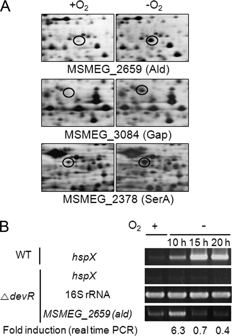
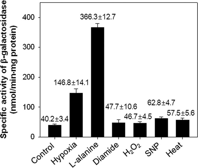
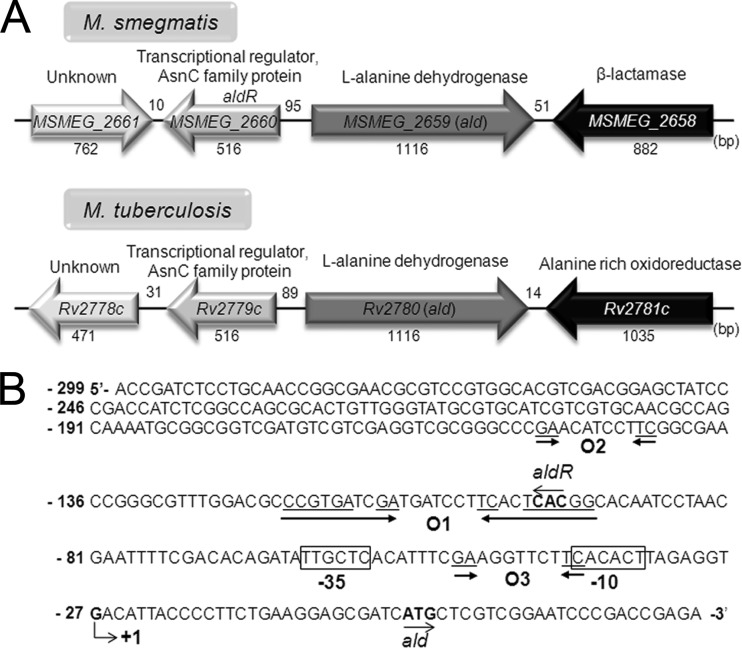
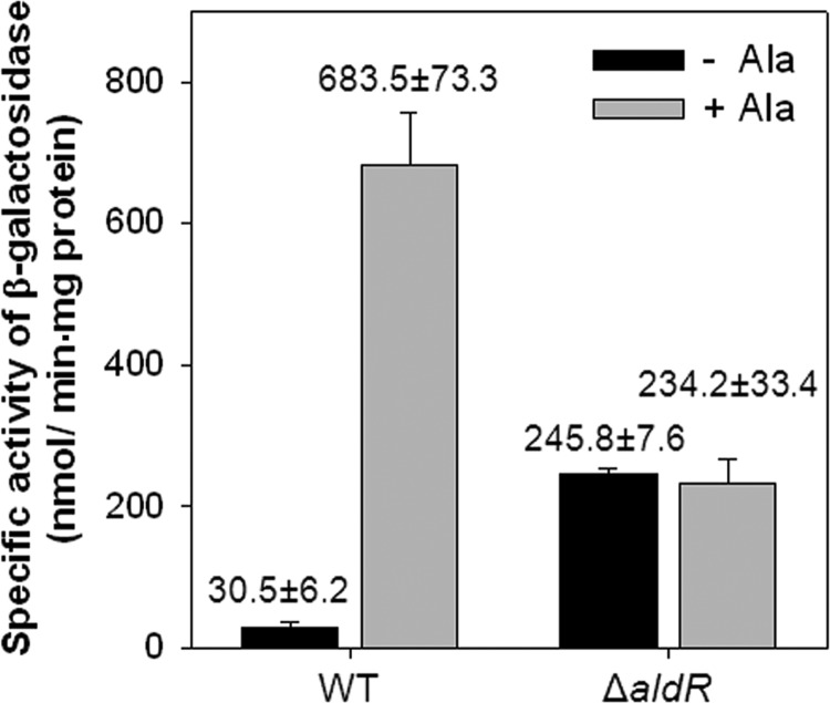
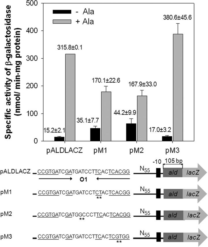
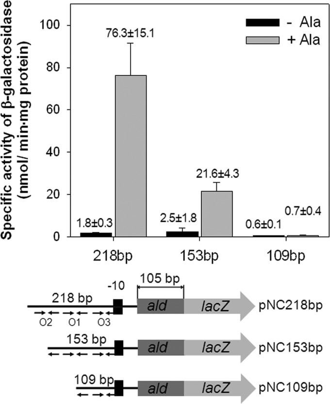
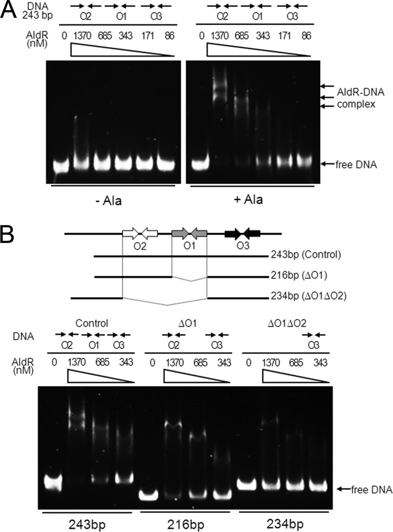
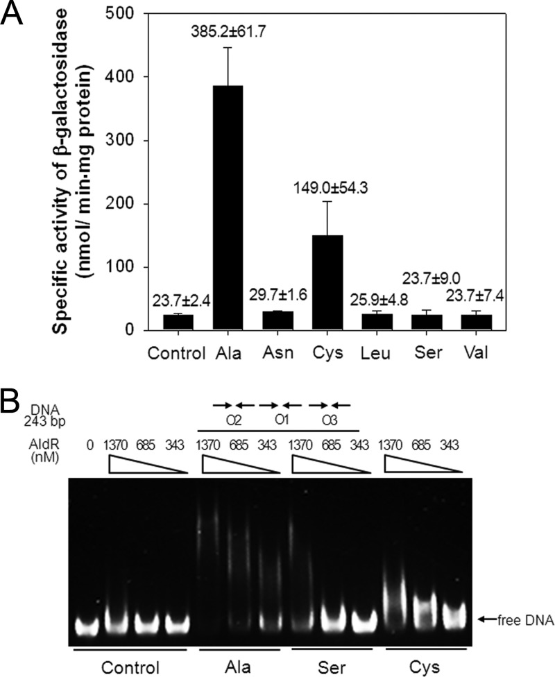
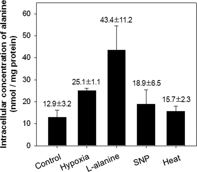
The regulatory gene aldR was identified 95 bp upstream of the ald gene encoding L-alanine dehydrogenase in Mycobacterium smegmatis. The AldR protein shows sequence similarity to the regulatory proteins of the Lrp/AsnC family. Using an aldR deletion mutant, we demonstrated that AldR serves as both activator and repressor for the regulation of ald gene expression, depending on the presence or absence of L-alanine. The purified AldR protein exists as a homodimer in the absence of L-alanine, while it adopts the quaternary structure of a homohexamer in the presence of L-alanine. The binding affinity of AldR for the ald control region was shown to be increased significantly by L-alanine. Two AldR binding sites (O1 and O2) with the consensus sequence GA-N₂-ATC-N₂-TC and one putative AldR binding site with the sequence GA-N₂-GTT-N₂-TC were identified upstream of the ald gene. Alanine and cysteine were demonstrated to be the effector molecules directly involved in the induction of ald expression. The cellular level of L-alanine was shown to be increased in M. smegmatis cells grown under hypoxic conditions, and the hypoxic induction of ald expression appears to be mediated by AldR, which senses the intracellular level of alanine.


Figures

Fig 1
Fig 1
Identification of the ald gene whose expression was upregulated in a ΔdevR mutant strain of M. smegmatis grown under hypoxic conditions. (A) Comparative 2D electrophoresis analysis of soluble proteins of the M. smegmatis ΔdevR strain grown under hypoxic (−O2) or aerobic (+O2) conditions for 20 h. The spots indicated by the open circles represent three proteins whose synthesis was increased more than 5-fold under hypoxic conditions compared to their levels under aerobic conditions. By means of MALDI-TOF MS, the proteins were identified as MSMEG_2659 (Ald), with a molecular mass of 38 kDa and pI of 5.9, MSMEG_3084 (Gap), with a molecular mass of 32 kDa and pI of 5.2, and MSMEG_2378 (SerA), with a molecular mass of 51 kDa and pI of 4.9. (B) Determination of the transcription levels of ald under hypoxic conditions. The wild-type (WT) and ΔdevR strains were grown either aerobically (+O2) or under hypoxic conditions (−O2) for 10, 15, and 20 h. The expression levels of ald and 16S rRNA genes were determined by RT-PCR. The hspX gene, whose expression was known to be induced by DevR under hypoxic conditions, was included in the experiment as a control. The levels of mRNA specific for ald were also determined by qRT-PCR and normalized to those of 16S rRNA. Fold induction of ald expression indicates the level of ald mRNA in hypoxic cultures relative to that in aerobic culture and is given below the RT-PCR results.
Fig 2
Fig 2
Expression of ald in the M. smegmatis ΔdevR strain exposed to various conditions. The M. smegmatis ΔdevR strain, carrying the ald::lacZ transcriptional fusion plasmid pALDLACZ, was grown either aerobically (control), under hypoxic conditions for 10 h (hypoxia), or exposed to various conditions as described in Materials and Methods. Cell-free crude extracts were used to determine β-galactosidase activity. The β-galactosidase activity is expressed as nanomoles per minute per milligram of protein. All values provided are the averages of the results from two independent determinations. Error bars indicate the deviations from the means.
Fig 3
Fig 3
Genetic organization of the ald locus and the putative cis-acting elements for ald expression. (A) The ald genes and their flanking genes in M. smegmatis mc2155 and M. tuberculosis H37Rv. The genes of the putative transcriptional regulators, MSMEG_2660 (aldR) and Rv2779c, are divergently located upstream of the ald genes. The lengths of genes and intergenic regions are given as the nucleotide numbers below and above their names, respectively. (B) The transcription start site (+1) of the ald gene of M. smegmatis was previously reported to be a guanosine residue that is located 27 nucleotides upstream of the start codon of ald (2). The −10 and −35 regions of the putative promoter for ald deduced from the transcription start point are boxed. The putative AldR binding site (O1), which shows an interrupted inverted repeat sequence ( CCGTGAN2GA-N7-TCN2TCACGG ), is marked by two head-facing arrows. The O2 and O3 sequences (GA-N7-TC) exhibiting partial sequence similarity to O1 are indicated by two arrows. The start codons of ald and aldR are highlighted in boldface and by the arrows indicating the transcriptional direction. The numbers on the left of the nucleotide sequence indicate the positions of the leftmost nucleotides relative to the ald gene.
Fig 4
Fig 4
Expression of the ald gene in the wild-type and ΔaldR mutant strains of M. smegmatis. M. smegmatis wild-type (WT) and ΔaldR strains containing the ald::lacZ transcriptional fusion plasmid (pALDLACZ) were grown aerobically in 7H9-glucose medium to an OD600 of 0.5 to 0.6. Following the addition of 25 mM l-alanine to the cultures, the strains were further grown for 1 h (+Ala). For the control, the M. smegmatis strains were grown aerobically without the addition of l-alanine (−Ala). The ald promoter activities were measured by determining the β-galactosidase activity. All values provided are the averages of the results from two independent determinations. Error bars indicate the deviations from the means.
Fig 5
Fig 5
Effect of base substitution mutations within the O1 site on ald expression. The ald promoter activities were determined by using the ald::lacZ transcriptional fusions with point mutations in the O1 region (pM1, pM2, and pM3), which are derivatives of pALDLACZ. Transition mutations within the O1 site are indicated by the asterisks. As the positive control, pALDLACZ was included in the experiment. M. smegmatis wild-type strains harboring the transcriptional fusion plasmids were grown aerobically in the presence (+Ala) or absence (−Ala) of l-alanine, and β-galactosidase activities were determined. All values provided are the averages of the results from two independent determinations. Error bars indicate the deviations from the means.
Fig 6
Fig 6
Promoter activities of the serially deleted upstream regions of ald in M. smegmatis. The ald promoter activities were determined by using ald::lacZ transcriptional fusions with 5′-nested deletions of the ald upstream region. The DNA fragments cloned into pNC consist of a shared 105-bp 5′ portion of ald and ald upstream regions in the different lengths denoted above the schematic diagrams (218, 153, and 109 bp). The relative positions and the presence or absence of the O1, O2, and O3 sites in the transcriptional fusions are indicated by the arrows. M. smegmatis wild-type strains harboring the transcriptional fusions were grown aerobically in the presence (+Ala) or absence (−Ala) of l-alanine, and β-galactosidase activities were determined. All values provided are the averages of results from two independent determinations. Error bars indicate the deviations from the means.
Fig 7
Fig 7
Binding of purified AldR to the ald control region. (A) The 243-bp DNA fragment (10 ng, corresponding to 6.7 nM) encompassing the O1, O2, and O3 sequences was incubated with various concentrations of purified AldR in the presence (+Ala) or absence (−Ala) of 20 mM l-alanine. The concentrations of AldR used are given above the lanes. Native PAGE was run in the presence of 83 mM Tris-borate buffer (pH 8.3) containing 1 mM EDTA, and the gels were stained with SYBR green EMSA gel staining solution. The arrows indicate bands of free DNA and retarded AldR-DNA complexes. (B) Ten ng of each of the DNA fragments (10 ng corresponds to 6.7 nM for control, 7.5 nM for ΔO1, and 6.9 nM for ΔO1O2), which contain O1, O2, and O3 (control), both O2 and O3 (ΔO1), and only O3 (ΔO1O2), were incubated with various concentrations of purified AldR in the presence of 20 mM l-alanine and subjected to native PAGE.
Fig 8
Fig 8
Effect of various amino acids on ald expression and binding of AldR to the ald control region. (A) M. smegmatis wild-type strain containing the ald::lacZ transcriptional fusion plasmid pALDLACZ was grown aerobically in 7H9 glucose medium to an OD600 of 0.5 to 0.6. Following the addition of 25 mM l-amino acids, the strain was further grown for an additional 1 h. The ald promoter activities were measured by determining the β-galactosidase activity. All values provided are the averages of results from two independent determinations. Error bars indicate the deviations from the means. (B) EMSA with the 243-bp DNA fragment (6.7 nM) encompassing O1, O2, and O3 and purified AldR in the presence of 20 mM l-alanine (Ala), l-serine (Ser), or l-cysteine (Cys). The concentrations of AldR used are given above the lanes. Native PAGE was run in the presence of 83 mM Tris-borate buffer (pH 8.3) containing 1 mM EDTA, and the gels were stained with SYBR green EMSA gel staining solution.
Fig 9
Fig 9
Determination of the intracellular alanine concentration in the M. smegmatis ΔdevR mutant strain grown under various conditions. To measure the intracellular alanine levels, the M. smegmatis ΔdevR mutant strain was grown either aerobically (control), under hypoxic conditions for 10 h (hypoxia), or exposed to various conditions (l-alanine, SNP, and 45°C heat) as described in Materials and Methods. The concentration of alanine in M. smegmatis ΔdevR cells was determined by spectrophotometrically measuring the amounts of NADH produced in a B. subtilis l-alanine dehydrogenase-linked enzyme assay. All values provided are the averages of the results from two independent determinations. Error bars indicate deviations from the means.

References

    1. Chen JM, Alexander DC, Behr MA, Liu J. 2003. Mycobacterium bovis BCG vaccines exhibit defects in alanine and serine catabolism. Infect. Immun. 71: 708– 716 - PMC - PubMed
    1. Feng Z, Caceres NE, Sarath G, Barletta RG. 2002. Mycobacterium smegmatis L-alanine dehydrogenase (Ald) is required for proficient utilization of alanine as a sole nitrogen source and sustained anaerobic growth. J. Bacteriol. 184: 5001– 5010 - PMC - PubMed
    1. Giffin MM, Modesti L, Raab RW, Wayne LG, Sohaskey CD. 2012. ald of Mycobacterium tuberculosis encodes both the alanine dehydrogenase and the putative glycine dehydrogenase. J. Bacteriol. 194: 1045– 1054 - PMC - PubMed
    1. Hutter B, Dick T. 1998. Increased alanine dehydrogenase activity during dormancy in Mycobacterium smegmatis. FEMS Microbiol. Lett. 167: 7– 11 - PubMed
    1. Betts JC, Lukey PT, Robb LC, McAdam RA, Duncan K. 2002. Evaluation of a nutrient starvation model of Mycobacterium tuberculosis persistence by gene and protein expression profiling. Mol. Microbiol. 43: 717– 731 - PubMed

Publication types

MeSH terms

LinkOut - more resources