. 2013 Aug;195(16):3629-39.
doi: 10.1128/JB.00347-13. Epub 2013 Jun 7.

Large linear plasmids of Borrelia species that cause relapsing fever

Affiliations

Large linear plasmids of Borrelia species that cause relapsing fever

Shelley Campeau Miller et al. J Bacteriol. 2013 Aug.

Abstract

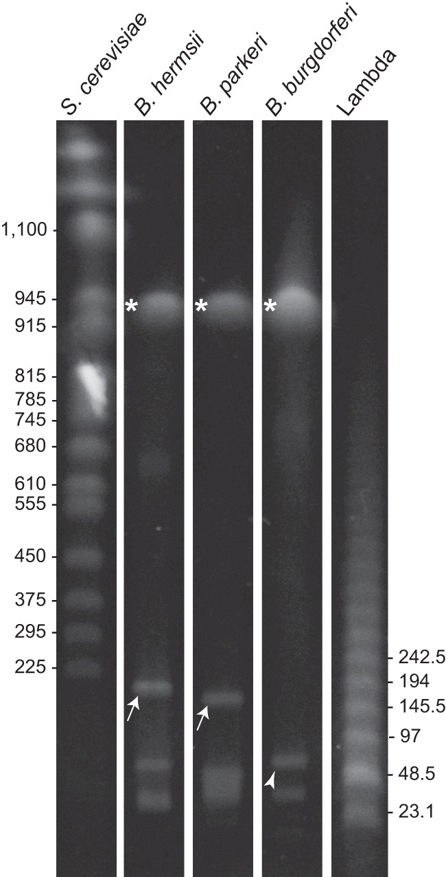
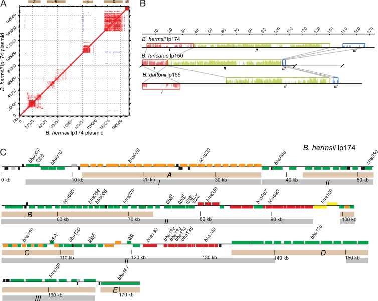
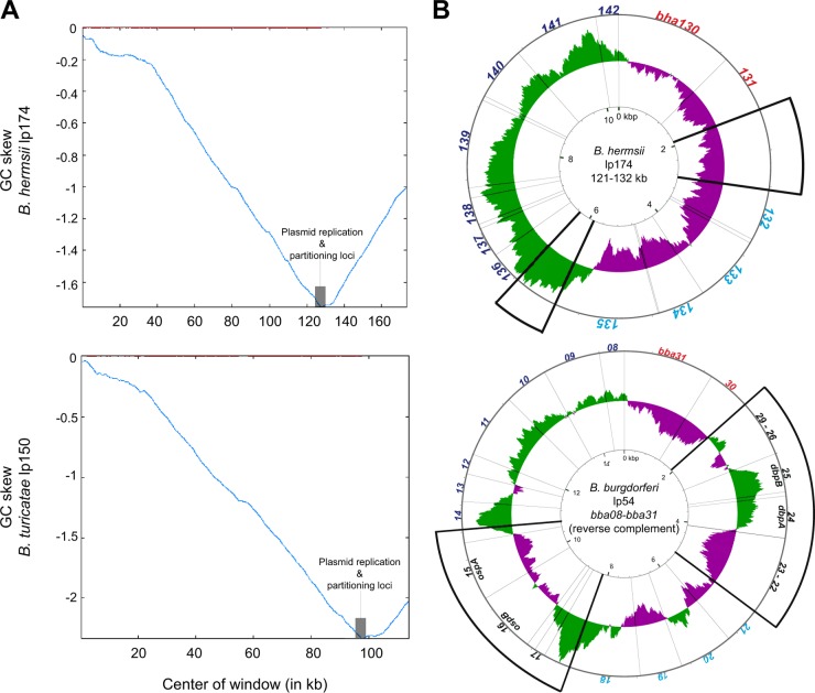
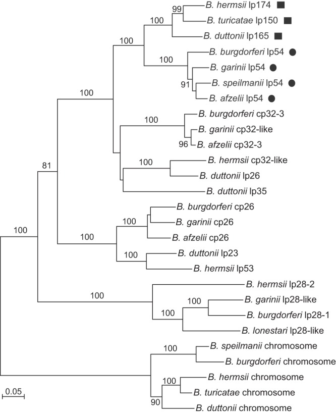
Borrelia species of relapsing fever (RF) and Lyme disease (LD) lineages have linear chromosomes and both linear and circular plasmids. Unique to RF species, and little characterized to date, are large linear plasmids of ∼160 kb, or ∼10% of the genome. By a combination of Sanger and next-generation methods, we determined the sequences of large linear plasmids of two New World species: Borrelia hermsii, to completion of its 174-kb length, and B. turicatae, partially to 114 kb of its 150 kb. These sequences were then compared to corresponding sequences of the Old World species B. duttonii and B. recurrentis and to plasmid sequences of LD Borrelia species. The large plasmids were largely colinear, except for their left ends, about 27 kb of which was inverted in New World species. Approximately 60% of the B. hermsii lp174 plasmid sequence was repetitive for 6 types of sequence, and half of its open reading frames encoded hypothetical proteins not discernibly similar to proteins in the database. The central ∼25 kb of all 4 linear plasmids was syntenic for orthologous genes for plasmid maintenance or partitioning in Borrelia species. Of all the sequenced linear and circular plasmids in Borrelia species, the large plasmid's putative partition/replication genes were most similar to those of the 54-kb linear plasmids of LD species. Further evidence for shared ancestry was the observation that two of the hypothetical proteins were predicted to be structurally similar to the LD species' CspA proteins, which are encoded on the 54-kb plasmids.


Figures

Fig 1
Fig 1
Pulsed-field gel of total DNAs of B. hermsii, B. parkeri, and B. burgdorferi. The large plasmids of B. hermsii and B. parkeri are indicated by arrows and the 54-kb linear plasmid of B. burgdorferi by an arrowhead. Asterisks indicate the linear chromosomes of all species. The sizes (in kb) of selected Saccharomyces cerevisiae chromosomes and lambda DNA concatemers are shown on the left and right, respectively.
Fig 2
Fig 2
Organization of repetitive DNA, colinear sequence, and individual open reading frames of relapsing fever Borrelia species. (A) Dot matrix plot of B. hermsii lp174 versus itself. Lines shown are similarities on direct strands (red) or on opposite strands (blue). Sequences were aligned using the default settings of the MAFFT alignment program. Regions of repetitive DNA are labeled A to E above the plot. (B) Schematic picture of alignment of the large linear plasmid sequences of B. hermsii, B. turicatae, and B. duttonii. Regions of contiguously homologous sequence are represented by colored local colinear blocks (LCBs I to III). An LCB is defined as a conserved segment of sequence that appears to be internally free from genome rearrangements relative to the reference sequence. Gray lines bound the orthologous LCBs. Similarity profiles (vertical lines within the LCBs) whose heights correspond to average level of sequence similarity are shown in each LCB. LCBs below the center line represent sequence in the opposite orientation relative to the reference sequence, that of B. hermsii lp174. Nucleotide positions (in kb) are indicated above the alignment. (C) Physical map of B. hermsii lp174 (accession no. HM008709) oriented left to right. Genes on the positive strand are indicated by boxes above the center line, and those on the negative strand are below the centerline. Beige shaded boxes (A to E) correspond to regions of repetitive DNA shown in panel A. Gray shaded boxes (I to III) correspond to LCBs shown in panel B. Selected genes previously mapped to the large plasmids are underlined: fhbA (bha008), nrdF (bha075), nrdE (bha076), nrdI (bha077), thyX (bha078), bipA (bha123), and alp (bha0128). Newly identified genes discussed in the text are also indicated: bha064, bha065, and the plasmid replication and partition loci (bha132 to -135). Colored ORFs represent homologs to the LD species P35 antigen (region A) or to PFam99 (orange), B. burgdorferi lp54 (red), B. burgdorferi cp26 (yellow), ORFs unique and conserved to RF species (green), or ORFs unique to a single species (black). Locus tags or gene names are indicated above the corresponding ORF.
Fig 3
Fig 3
GC skew analysis of Borrelia plasmid sequences. (A) GC skew diagrams of B. hermsii lp174 and B. turicatae partial lp150 sequences. Shaded gray boxes indicate the plasmid replication and partitioning loci. Red lines at the top indicate the window and the point at which the skew shifts. Total GC skew was calculated with a sliding window size of 500 bp and a step size of 50 bp. (B) Circular G-C skew representation of orthologous regions from B. hermsii lp174 and B. burgdorferi lp54. Deviation from the average GC skew of the entire sequence is represented by green lines (above average) or purple lines (below average). Radiating lines originating from the center circle indicate ORF boundaries. Orthologous ORFs are noted by the same font colors (red, cyan, or blue), with thick diagonal lines enclosing regions of nonhomologous sequence (ORFs indicated in black). GC skew was calculated with a 500-bp window and a step size of 1 bp (75).
Fig 4
Fig 4
Phylogram of codon-aligned nucleotide sequences for selected PFam32 orthologs from different Borrelia replicons. Genes from the RF species large plasmids (squares) and LD species lp54 plasmids (circles) are indicated. The replicons and accession numbers used for each species and strain are provided in Materials and Methods. Nodes with bootstrap values of >70% support by neighbor-joining distance criteria from 10,000 replicates are indicated. The scale bar represents nucleotide substitutions per site.
Fig 5
Fig 5
Gene expression analysis of bha064. Quantitative real-time PCR analysis of bha064 cDNA copies normalized to copies of the constitutively expressed flaB for B. hermsii grown at 37°C (gray bars) in the blood of immunodeficient mice or in culture or grown at 23°C (black bars) in culture is shown. Ninety-five percent confidence intervals are shown for each condition. Asterisks above lines indicate significant differences as measured by the t test (P < 0.001).

References

    1. Levin BR, Bergstrom CT. 2000. Bacteria are different: observations, interpretations, speculations, and opinions about the mechanisms of adaptive evolution in prokaryotes. Proc. Natl. Acad. Sci. U. S. A. 97:6981–6985 - PMC - PubMed
    1. Ferdows MS, Barbour AG. 1989. Megabase-sized linear DNA in the bacterium Borrelia burgdorferi, the Lyme disease agent. Proc. Natl. Acad. Sci. U. S. A. 86:5969–5973 - PMC - PubMed
    1. Casjens S, Huang WM. 1993. Linear chromosomal physical and genetic map of Borrelia burgdorferi, the Lyme disease agent. Mol. Microbiol. 8:967–980 - PubMed
    1. Plasterk RH, Simon MI, Barbour AG. 1985. Transposition of structural genes to an expression sequence on a linear plasmid causes antigenic variation in the bacterium Borrelia hermsii. Nature 318:257–263 - PubMed
    1. Crespi M, Messens E, Caplan AB, van Montagu M, Desomer J. 1992. Fasciation induction by the phytopathogen Rhodococcus fascians depends upon a linear plasmid encoding a cytokinin synthase gene. EMBO J. 11:795–804 - PMC - PubMed

Publication types

Substances

LinkOut - more resources