. 2013 Jun 4;8(6):e64992.
doi: 10.1371/journal.pone.0064992. Print 2013.

Altered functionality of anti-bacterial antibodies in patients with chronic hepatitis C virus infection

Affiliations

Altered functionality of anti-bacterial antibodies in patients with chronic hepatitis C virus infection

Anne Lamontagne et al. PLoS One. .

Abstract

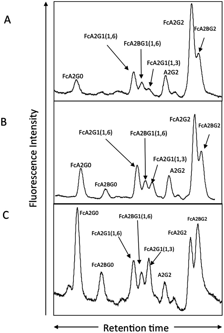
Background: Using comparative glycoproteomics, we have previously identified a glycoprotein that is altered in both amount and glycosylation as a function of liver cirrhosis. The altered glycoprotein is an agalactosylated (G0) immunoglobulin G molecule (IgG) that recognizes the heterophilic alpha-gal epitope. Since the alpha gal epitope is found on gut enterobacteria, it has been hypothesized that anti-gal antibodies are generated as a result of increased bacterial exposure in patients with liver disease.

Methods: The N-linked glycosylation of anti-gal IgG molecules from patients with fibrosis and cirrhosis was determined and the effector function of anti-bacterial antibodies from over 100 patients examined. In addition, markers of microbial exposure were determined.

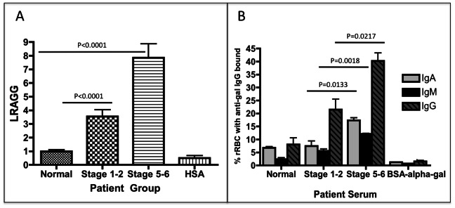
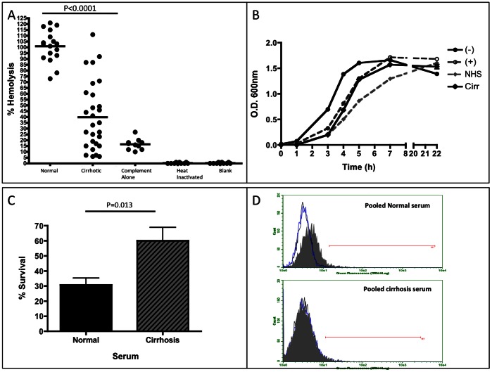
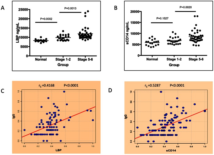
Results: Surprisingly, the subset of agalactosylated anti-gal antibodies described here, was impaired in their ability to mediate complement mediated lysis and inhibited the complement-mediated destruction of common gut bacteria. In an analysis of serum from more than 100 patients with liver disease, we have shown that those with increased levels of this modified anti-gal antibody had increased levels of markers of bacterial exposure.

Conclusions: Anti-gal antibodies in patients with liver cirrhosis were reduced in their ability to mediate complement mediated lysis of target cells. As bacterial infection is a major complication in patients with cirrhosis and bacterial products such as LPS are thought to play a major role in the development and progression of liver fibrosis, this finding has many clinical implications in the etiology, prognosis and treatment of liver disease.


Conflict of interest statement

Competing Interests: Author Julie Hafner is affiliated to Immunotope Inc. There are no patents, products in development or marketed products to declare. This does not alter the authors' adherence to all the PLOS ONE policies on sharing data and materials.

Figures

Figure 1
Figure 1. The glycosylation of anti-gal IgG is altered with the development of fibrosis and cirrhosis.
Anti-gal IgG was purified from a pool (n = 20) of healthy individuals, pooled ( = 20) fibrotic individuals (stage 1–2) or pooled (n = 20) late stage fibrotic (cirrhotic) individuals (stage 5–6). Glycan analysis of the N-linked glycans associated with the heavy chain from the healthy individuals (panel A), the fibrotic individuals (panel B) or the late stage fibrotic individuals (panel C). For structures presented in panels A-C: FcA2G0, core fucosylated (1,6) agalactosylated biantennary glycan; FcA2G0B, core fucosylated (1,6) agalactosylated biantennary glycan with a bisecting N-acetylglucosamine (GlcNac); FcA2G1 (1,6) core fucosylated (1,6) biantennary glycan with a single galactose residue on the 1,6 arm; FcA2G1B (1,6) core fucosylated (1,6) biantennary glycan with a single galactose residue on the 1,6 arm and a bisecting GlcNac; FcA2G1 (1,3), core fucosylated (1,6) biantennary glycan with a single galactose residue on the 1,3 arm; FcA2G2, core fucosylated biantennary N-glycan (FcA2G2); FcA2G2B, bisected core fucosylated biantennary N-glycan.
Figure 2
Figure 2. Increased lectin reactive anti-gal IgG from patients with increasing levels of liver fibrosis.
A) Compared to commercially purchased normal human sera, patients with limited liver fibrosis (stage 1–2) have a 3.5 (±1.4) fold increase in lectin reactive anti-gal IgG (LRAGG). More advanced (Stage 5–6) fibrosis patients have a 7.9 (±2.4) fold increase in LRAGG. The fold increase in LRAGG is statistically significant (P<0.001). When serum from more advanced fibrotic patients is used on plates coated with HSA alone and not HSA-alpha-gal, no signal is observed (HSA lane). B) The level of anti-gal IgA, IgM and IgG bound to target rabbit red blood cells as a function of liver fibrosis. There is a statistically significant increase in anti-gal IgA from commercially purchased normal human serum to serum from patients with advanced fibrosis (P = 0.0048); Anti-gal IgA also significantly increases from limited to advanced fibrosis (P = 0.0133). Anti-gal IgM significantly increases from control to limited fibrosis (P = 0.02) and from control to advanced fibrosis (P = 0.002); there is also a significant increase in anti-gal IgM from limited to advanced fibrosis (P = 0.0018). Anti-gal IgG is significantly elevated in advanced fibrosis compared to control (P = 0.0075) and also from limited to advanced fibrosis (P = 0.0217). 50 mM of BSA-alpha-gal can prevent binding of antibodies to RBCC. See text for more details. For panels A & B, samples size is Normal, n = 21; Stage 1–2, n = 22; and Cirrhosis, n = 39.
Figure 3
Figure 3. Anti-gal specific antibodies have poor complement mediated killing ability and are unable to induce phagocytosis of opsonized target cells.
A) Results from a hemoglobin release assay using serum from control patients and patients with cirrhosis. Compared to normal serum, serum from cirrhosis patients has an over 60% decrease in the capacity to induce complement mediated killing of target rRBCs. For panel A, sample size is: Normal, n = 20 and Cirrhotics, n = 20. As a control, complement alone was used to indicate the level of the alternative pathway (complement alone). In addition, if normal samples were heat inactivated or not treated with serum or complement, no lysis was observed. B) Results from a bactericidal assay using human serum show the growth pattern of bacteria alone (–), bacteria incubated with functional complement (+), bacteria with functional complement and normal human serum (NHS), or bacteria with functional complement and serum from a pool of 20 cirrhosis patients (Cirr). Error bars are indicated. C) Bacteria incubated with serum from a pool of 20 cirrhosis patients, in the presence of functional complement, show a significantly increased survival rate compared to those exposed to normal human serum (P = 0.013). Data normalized to those that did not receive serum addition. D) Results from an opsonization/phagocytosis assay. Bottom panel show target cells opsonized with serum from cirrhosis patients are not phagocytosed by monocytes, while top panel shows target cells opsonized with purchased normal serum are phagocytosed. Black peak represents monocytes alone, blue peak represents monocytes incubated with non-opsonized target cells, and gray filled peak represents opsonized target cells incubated with monocytes.
Figure 4
Figure 4. Peripheral markers of LPS exposure in patients with liver fibrosis correlate with the level of LRAGG.
A) There is a statistically significant increase in the level of LPS Binding Protein (LBP) detected in the sera of patients with mild liver fibrosis (P = 0.0002) and also as a function of liver cirrhosis (P = 0.0013). B) A similar increase is seen in soluble CD14 (sCD14) with the progression of liver fibrosis to liver cirrhosis (P = 0.0020). C) There is a direct correlation between LBP and LRAGG (rS = 0.4168, P<0.0001) and between sCD14 and LRAGG (rS = 0.5287, P<0.0001) (D). rS denotes the Spearman’s correlation coefficient. For panels A & B, samples size is Normal, n = 20; Stage 1–2, n = 21; and Stage 5–6 (Cirrhosis), n = 39.
Figure 5
Figure 5. Change in LRAGG in response to IFN Treatment in patients with liver fibrosis.
(A) The change in the level of LRAGG in patients who responded to IFN and cleared their HCV and had low LRAGG at start of treatment. As this figure shows, there is no significant change in patients who started with low levels of LRAGG in response to IFN therapy. (B) In contrast, those patients who responded to IFN and cleared their HCV and started had greater than 5-fold increase over normal at the start of treatment show a significant decrease following treatment (P<0.05) (C). However, patients who do not respond to IFN treatment show a significant increase (P<0.05) following treatment when they began with low levels (C) and stayed high when they started with greater than 5-fold increase in LRAGG over normal (D).
Figure 6
Figure 6. Markers of bacterial exposure change in response to IFN therapy in patients with liver fibrosis.
Patient sera were tested for sCD14 and LBP levels pre- and post- IFN therapy. In patients who respond to treatment, there is a statistically significant decrease in sCD14 following treatment (P<0.0001) while patients who do not respond show a significant increase following the course of treatment (P<0.0001) (B). There was no significant change LBP between treatment groups in patients who respond to IFN therapy (C), however patients who do not respond to treatment show a statistically significant increase from pre-treatment LBP levels (P<0.001) (D).

References

    1. Alter MJ (1997) Epidemiology of hepatitis C. Hepatology. 26: 62S–65S. - PubMed
    1. Hoofnagle JH (1997) Hepatitis C: the clinical spectrum of disease. Hepatology 26: 15S–20S. - PubMed
    1. Di Bisceglie AM (1997) Hepatitis C and hepatocellular carcinoma. Hepatology 26: 34S–38S. - PubMed
    1. Iredale JP (2007) Models of liver fibrosis: exploring the dynamic nature of inflammation and repair in a solid organ. J Clin Invest 117: 539–548. - PMC - PubMed
    1. Friedman SL (2007) A deer in the headlights: BAMBI meets liver fibrosis. Nat Med 13: 1281–1282. - PubMed

Publication types