Comparative Study
. 2013 Oct;174(1):161-71.
doi: 10.1111/cei.12155.

Whole blood stimulation with Toll-like receptor (TLR)-7/8 and TLR-9 agonists induces interleukin-12p40 expression in plasmacytoid dendritic cells in rhesus macaques but not in humans

Affiliations
Comparative Study

Whole blood stimulation with Toll-like receptor (TLR)-7/8 and TLR-9 agonists induces interleukin-12p40 expression in plasmacytoid dendritic cells in rhesus macaques but not in humans

G Koopman et al. Clin Exp Immunol. 2013 Oct.

Abstract

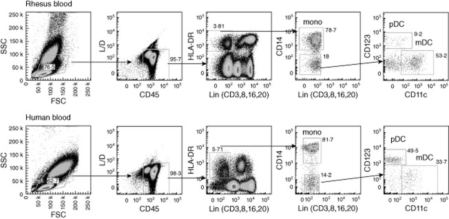
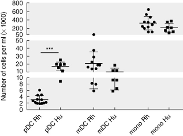
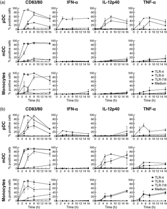
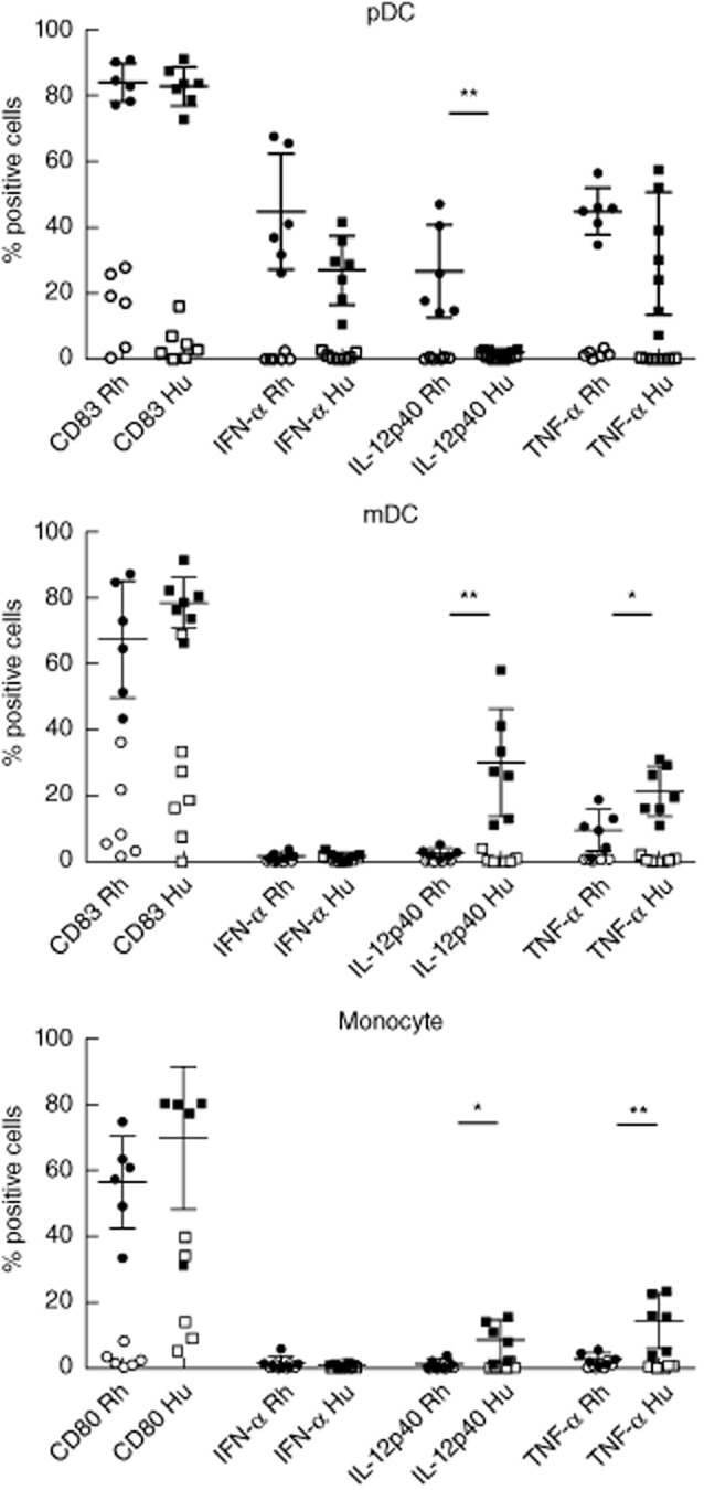
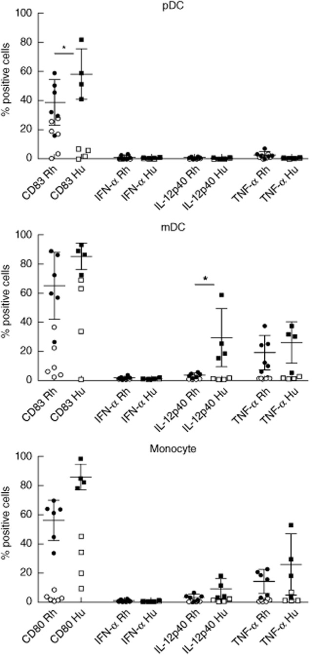
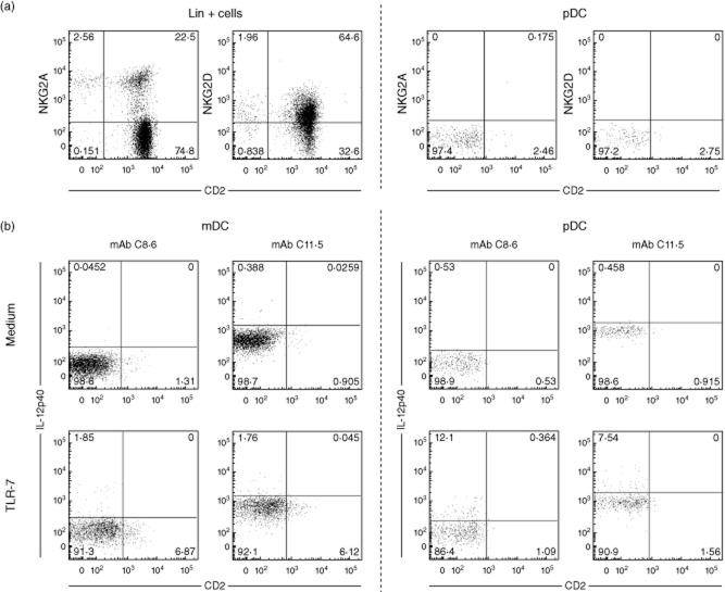
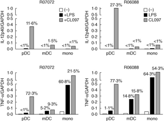
Macaques provide important animal models in biomedical research into infectious and chronic inflammatory disease. Therefore, a proper understanding of the similarities and differences in immune function between macaques and humans is needed for adequate interpretation of the data and translation to the human situation. Dendritic cells are important as key regulators of innate and adaptive immune responses. Using a new whole blood assay we investigated functional characteristics of blood plasmacytoid dendritic cells (pDC), myeloid dendritic cells (mDC) and monocytes in rhesus macaques by studying induction of activation markers and cytokine expression upon Toll-like receptor (TLR) stimulation. In a head-to-head comparison we observed that rhesus macaque venous blood contained relatively lower numbers of pDC than human venous blood, while mDC and monocytes were present at similar percentages. In contrast to humans, pDC in rhesus macaques expressed the interleukin (IL)-12p40 subunit in response to TLR-7/8 as well as TLR-9 stimulation. Expression of IL-12p40 was confirmed by using different monoclonal antibodies and by reverse transcription-polymerase chain reaction (RT-PCR). Both in humans and rhesus macaques, TLR-4 stimulation induced IL-12p40 expression in mDC and monocytes, but not in pDC. The data show that, in contrast to humans, pDC in macaques are able to express IL-12p40, which could have consequences for evaluation of human vaccine candidates and viral infection.

Keywords: Toll-like receptors (TLRs); animal models - non-human primates; cytokine/interleukin/chemokine receptors; dendritic cells (myeloid; monocyte-derived); plasmacytoid.


Figures

Fig. 1
Fig. 1
Analysis of rhesus versus human peripheral blood dendritic cell (DC) subsets. For analysis of monocytes and DC in rhesus macaques (top row) and humans (bottom row), an extensive lymphogate is drawn in the forward-scatter (FSC)/side-scatter plot (SSC), encompassing all lymphocyte, monocyte and DC subsets. Subsequently, cells negative for live/dead (L/D) stain and positive for CD45 are gated and expression of lineage markers (combination of CD3, CD8, CD16, CD20) is plotted against human leucocyte antigen D-related (HLA-DR). Within this plot the Lin, HLA-DR+ cells are selected and expression of CD14 is used to identify the monocytes. The CD14 cells are then divided into a CD11c+/CD123 mDC and a CD11c/CD123+ pDC subset. Figures in the graphs indicate per gate the number of cells as a percentage of the parental population. The relative difference in plasmacytoid dendritic cell (pDC) percentages between the rhesus and human sample is striking.
Fig. 2
Fig. 2
Absolute number of plasmacytoid dendritic cells (pDC), myeloid dendritic cells (mDC) and monocytes in rhesus (n = 12) and human (n = 7) peripheral blood. Indicated is the number of cells (×1000)/ml blood. ***P < 0·001.
Fig. 3
Fig. 3
Kinetics of Toll-like receptor (TLR)-7/-8-, TLR-9- and TLR-4-induced CD83 or CD80 and cytokine expression in rhesus (a) and human (b) peripheral blood plasmacytoid dendritic cells (pDC), myeloid dendritic cells (mDC) and monocytes. For each subset the percentage of cells expressing CD83 (pDC and mDC) or CD80 (monocytes), interferon (IFN)-α, interleukin (IL)-12p40 or tumour necrosis factor (TNF)-α after 3, 5, 8 or 16 h stimulation with TLR-7/8 (1 μg/ml CL097), TLR-9 [5 μM class C cytosine–phosphate–guanosine (CpG-C)] or Toll-like receptor (TLR)-4 (1 μg/ml lipopolysaccharide) is shown. Golgiplug was added after 1, 1, 2 and 2 h of incubation, respectively. Indicated is mean expression with error of two independent experiments.
Fig. 4
Fig. 4
Toll-like receptor (TLR)-7/-8-induced CD83 or CD80 and cytokine expression in rhesus versus human peripheral blood plasmacytoid dendritic cells (pDC), myeloid dendritic cells (mDC) and monocytes. For each subset the percentage of cells expressing CD83 (pDC and mDC) or CD80 (monocytes), interferon (IFN)-α, interleukin (IL)-12p40 or tumour necrosis factor (TNF)-α after 8 h stimulation with 1 μg/ml CL097 is shown (filled symbols); background expression as observed in cultured non-stimulated cells (open symbols) was not subtracted. *P < 0·05; **P < 0·01.
Fig. 5
Fig. 5
Toll-like receptor (TLR)-9-induced CD83 and cytokine expression in rhesus (n = 6) versus human (n = 2) peripheral blood plasmacytoid dendritic cells (pDC), myeloid dendritic cells (mDC) and monocytes. For each subset the percentage of cells expressing CD83 (pDC and mDC), interferon (IFN)-α, interleukin (IL)-12p40 or tumour necrosis factor (TNF)-α after 8 h stimulation with 5 μM class C cytosine–phosphate–guanosine (CpG-C) (M362) is shown (filled symbols); background expression as observed in cultured non-stimulated cells (open symbols) was not subtracted; n.d.: not done.
Fig. 6
Fig. 6
Toll-like receptor (TLR)-4-induced CD83 and cytokine expression in rhesus (n = 6) versus human (n = 4) peripheral blood plasmacytoid dendritic cells (pDC), myeloid dendritic cells (mDC) and monocytes. For each subset the percentage of cells expressing CD83 (pDC and mDC) or CD80 (monocytes), interferon (IFN)-α, interleukin (IL)-12p40 or tumour necrosis factor (TNF)-α after 8 h stimulation with 1 μg/ml lipopolysaccharide is shown (filled symbols); background expression as observed in cultured non-stimulated cells (open symbols) was not subtracted. *P < 0·05.
Fig. 7
Fig. 7
Phenotypic analysis of rhesus plasmacytoid dendritic cells (pDC) and induction of interleukin (IL)-12p40 expression. (a) Shown is expression of CD2 versus natural killer (NK) type G2A (NKG2A) or NKG2D on lineage positive cells (two left graphs) or pDC [lineage/human leucocyte antigen-D-related (HLA-DR)+/CD11c/CD123+) (two right graphs). (b) Expression of IL-12p40 (vertical axis) in mDC and pDC in unstimulated (medium) versus Toll-like receptor (TLR)-7/-8 (CL097)-stimulated cells, detected with either monoclonal antibody (mAb) C8·6 or mAb 11·5.
Fig. 8
Fig. 8
Interleukin (IL)-12p40 and tumour necrosis factor (TNF)-α mRNA expression in Toll-like receptor (TLR)-4 (lipopolysaccharide) and TLR-7/8 (CL097) stimulated purified rhesus macaque plasmacytoid dendritic cells (pDC), myeloid dendritic cells (mDC) and monocytes. Shown are expression levels of IL-12p40 (top graphs) and TNF-α (bottom graphs) relative to glyceraldehyde 3-phosphate dehydrogenase (GAPDH) in pDC, mDC and monocytes as well as percentage of positive cells as detected by fluorescence activated cell sorter (FACS) analysis on the same cells (number at top of bars). Results from two individual rhesus macaques are shown.

References

    1. Bontrop RE. Non-human primates: essential partners in biomedical research. Immunol Rev. 2001;183:5–9. - PubMed
    1. Banchereau J, Steinman RM. Dendritic cells and the control of immunity. Nature. 1998;392:245–252. - PubMed
    1. Navarro-Sanchez E, Altmeyer R, Amara A, et al. Dendritic-cell-specific ICAM3-grabbing non-integrin is essential for the productive infection of human dendritic cells by mosquito-cell-derived dengue viruses. EMBO Rep. 2003;4:723–728. - PMC - PubMed
    1. Lozach PY, Lortat-Jacob H, de Lacroix de Lavalette A, et al. DC-SIGN and L-SIGN are high affinity binding receptors for hepatitis C virus glycoprotein E2. J Biol Chem. 2003;278:20358–20366. - PubMed
    1. Geijtenbeek TB, Kwon DS, Torensma R, et al. DC-SIGN, a dendritic cell-specific HIV-1-binding protein that enhances trans-infection of T cells [see comments] Cell. 2000;100:587–597. - PubMed

Publication types

MeSH terms