

Polymicrogyria-associated epilepsy: a multicenter phenotypic study from the Epilepsy Phenome/Genome Project
- PMID: 23750890
- PMCID: PMC3851304
- DOI: 10.1111/epi.12238
Polymicrogyria-associated epilepsy: a multicenter phenotypic study from the Epilepsy Phenome/Genome Project
Abstract
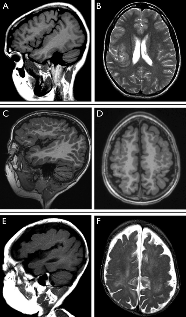
Purpose: Polymicrogyria (PMG) is an epileptogenic malformation of cortical development. We describe the clinical epilepsy and imaging features of a large cohort with PMG-related epilepsy.
Methods: Participants were recruited through the Epilepsy Phenome/Genome Project, a multicenter collaborative effort to collect detailed phenotypic data on individuals with epilepsy. We reviewed phenotypic data from participants with epilepsy and PMG.
Key findings: We identified 87 participants, 43 female and 44 male, with PMG and epilepsy. Median age of seizure onset was 3 years (range <1 month to 37 years). Most presented with focal epilepsy (87.4%), some in combination with seizures generalized from onset (23.0%). Focal seizures with dyscognitive features were most common (54.3%). Of those presenting with generalized seizure types, infantile spasms were most prevalent (45.2%). The most common topographic pattern was perisylvian PMG (77.0%), of which the majority was bilateral (56.7%). Generalized PMG presented with an earlier age of seizure onset (median age of 8 months) and an increased prevalence of developmental delay prior to seizure onset (57.1%). Of the unilateral, and asymmetric bilateral groups where PMG was more involved in one hemisphere, the majority (71.4%) of participants had seizures that lateralized to the same hemisphere as the PMG or the hemisphere with greater involvement.
Significance: Participants with PMG had both focal and generalized onset of seizures. Our data confirm the involvement of known topographic patterns of PMG and suggest that more extensive distributions of PMG present with an earlier age of seizure onset and increased prevalence of developmental delay prior to seizure onset.
Keywords: Epilepsy; Epilepsy Phenome/Genome Project; Perisylvian; Polymicrogyria.
Wiley Periodicals, Inc. © 2013 International League Against Epilepsy.
Figures
References
-
- Baykan-Kurt B, Sarp A, Gokyigit A, Tuncay R, Caliskan A. A clinically recognizable neuronal migration disorder: congenital bilateral perisylvian syndrome. Case report with long-term clinical and EEG follow-up. Seizure. 1997;6:487–493. - PubMed
-
- Berg AT, Berkovic SF, Brodie MJ, Buchhalter J, Cross JH, van Emde Boas W, Engel J, French J, Glauser TA, Mathern GW, Moshe SL, Nordli D, Plouin P, Scheffer IE. Revised terminology and concepts for organization of seizures and epilepsies: report of the ILAE Commission on Classification and Terminology, 2005–2009. Epilepsia. 2010;51:676–685. - PubMed
-
- Brodtkorb E, Andersen K, Henriksen O, Myhr G, Skullerud K. Focal, continuous spikes suggest cortical developmental abnormalities. Clinical, MRI and neuropathological correlates. Acta Neurol Scand. 1998;98:377–385. - PubMed
-
- Brodtkorb E, Nilsen G, Smevik O, Rinck PA. Epilepsy and anomalies of neuronal migration: MRI and clinical aspects. Acta Neurol Scand. 1992;86:24–32. - PubMed
Publication types
MeSH terms
Grants and funding
LinkOut - more resources
Full Text Sources
Other Literature Sources
Medical
Molecular Biology Databases
