Review
. 2013 Jul;10(7):907-26.
doi: 10.1517/17425247.2013.808184. Epub 2013 Jun 11.

Targeted delivery of antibody-based therapeutic and imaging agents to CNS tumors: crossing the blood-brain barrier divide

Affiliations
Review

Targeted delivery of antibody-based therapeutic and imaging agents to CNS tumors: crossing the blood-brain barrier divide

Ann-Marie Chacko et al. Expert Opin Drug Deliv. 2013 Jul.

Erratum in

  • Expert Opin Drug Deliv. 2013 Oct;10(10):1463

Abstract

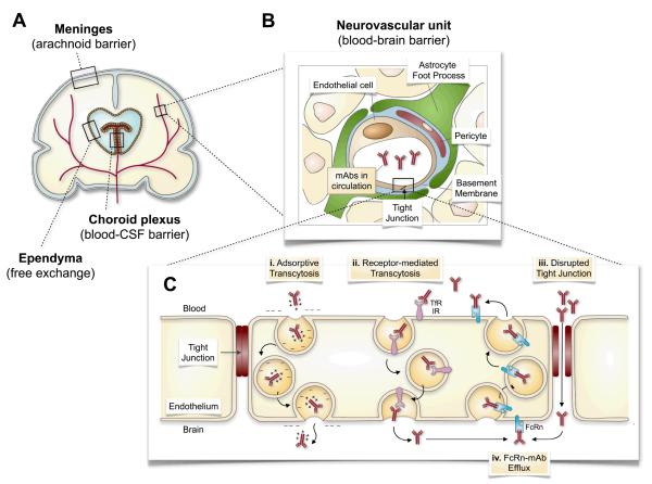
Introduction: Brain tumors are inherently difficult to treat in large part due to the cellular blood-brain barriers (BBBs) that limit the delivery of therapeutics to the tumor tissue from the systemic circulation. Virtually no large molecules, including antibody-based proteins, can penetrate the BBB. With antibodies fast becoming attractive ligands for highly specific molecular targeting to tumor antigens, a variety of methods are being investigated to enhance the access of these agents to intracranial tumors for imaging or therapeutic applications.

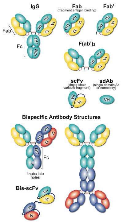
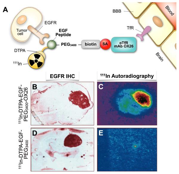
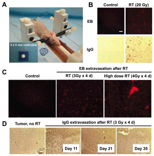
Areas covered: This review describes the characteristics of the BBB and the vasculature in brain tumors, described as the blood-brain tumor barrier (BBTB). Antibodies targeted to molecular markers of central nervous system (CNS) tumors will be highlighted, and current strategies for enhancing the delivery of antibodies across these cellular barriers into the brain parenchyma to the tumor will be discussed. Noninvasive imaging approaches to assess BBB/BBTB permeability and/or antibody targeting will be presented as a means of guiding the optimal delivery of targeted agents to brain tumors.

Expert opinion: Preclinical and clinical studies highlight the potential of several approaches in increasing brain tumor delivery across the BBB divide. However, each carries its own risks and challenges. There is tremendous potential in using neuroimaging strategies to assist in understanding and defining the challenges to translating and optimizing molecularly targeted antibody delivery to CNS tumors to improve clinical outcomes.


Figures

Figure 1
Figure 1
Routes for antibody delivery across the blood-brain barrier (BBB). (A) The brain is comprised of multiple barriers that control access of administered agents into the brain parenchyma, including the BBB. (B) The BBB is comprised of a neurovascular unit that consists of pericytes, astrocytes, and neurons surrounding brain capillary endothelial cells (and basement membrane). (C) Expansion of the endothelial lining of the BBB shows that intact tight junctions in the normal brain restrict the intercellular diffusion of mAb across the BBB into the brain. (i) Adsorptive transcytosis of cationized mAb can increase transcellular transport across the BBB by their interaction with naturally negatively charged plasma membrane. (ii) MAb engagement of the transferrin receptor (TfR) or the insulin receptor (IR), for example, can also increase mAb transport across the BBB by receptor-mediated transcytosis. (iii) The disruption of tight junctions by pharmacological (osmotic, biochemical) and mechanical (ultrasound, irradiation) can enhance paracellular extravasation of mAbs into the brain parenchyma. (iv) The neonatal Fc receptor (FcRn) serves as an efflux mechanism to remove Fc-containing mAb from the brain and return them to the systemic circulation. Figure 1A and 1B adapted with permission from Elsevier Ltd. (Copyright © 2008): Saunders, N.R. et al. Barriers in the brain: a renaissance? Trends in Neurosciences. 31(6): 279-286. Abbreviations: CSF, cerebrospinal fluid; mAb, monoclonal antibody; TfR, transferrin receptor; IR, insulin receptor; FcRn, neonatal Fc receptor.
Figure 2
Figure 2
Schematic representation of different antibody formats. An array of recombinant antibody formats were developed using combinations of antibody domains, joined either with linkers and/or disulfide bonds. Using molecular biology tools, antibody valency and specificity can be tailored into monovalent (sdAb (single domain Ab); Fab and Fab’ (fragment antigen binding), divalent (F(ab’)2) and even multivalent formats. Formats with antibody constant domains are highly stable, however, single chain antibody formats are popular due to their small size, expression and tissue penetration capabilities. Linker length can also influence the antibody behavior in terms of stability, flexibility and aggregation. A bispecific antibody is a recombinant antibody format that recognize two distinct antigens, using two IgG or scFv, linked together either chemically by covalent conjugation; or genetically with peptide linkers; or disulfide bonds.
Figure 3
Figure 3
A, Structure of dual targeted epidermal growth factor (EGF) chimeric peptide conjugated to rat transferrin receptor (TfR) targeted mAb OX26. The EGF is modified with diethylenetriamine pentaacetic acid (DTPA) to chelate the radioisotope 111In. The EGF is attached to a 3400-Da polyethylene glycol linker (PEG3400) terminated with a biotin moiety for conjugation to streptavidin (SA)-OX26 MAb. B, D. Brain sections of U87 human glioma tumor-bearing rats were stained immunocytochemically using an MAb to the human EGF receptor (EGF-R), which demonstrates expression of the EGF-R in brain tumor specimens. C, E. Ex vivo 111In-autoradiography demonstrates that the brain tumor can be imaged with the EGF chimeric peptide that can undergo transport across the BBB in vivo via antiTfR mAb OX26. (C) There is no imaging of the brain tumor when the non TfR-targeted EGF peptide radiopharmaceutical is administered, because the EGF does not cross the BBB alone (E). Adapted with permission from the American Association for Cancer Research (Copyright © 1999): Kurihara A and Pardridge WM. Imaging brain tumors by targeting peptide radiopharmaceuticals through the blood-brain barrier. Cancer Res. 1999; 59 (24), 6159-63.
Figure 4
Figure 4. Targeted radiation therapy disrupts brain tumor blood-brain barrier and permits greater penetration of Evans Blue dye and IgG
A, Cranial RT is delivered using a novel mouse restrainer housed within the Small Animal Radiation Research Platform (SARRP). RT is focused using a 5 × 5 mm collimator (inset). B, Healthy nude mice, mock-irradiated (“Control”, left column) or irradiated with 20 Gy in a single fraction to the right cerebral hemisphere (“RT”, right column), show BBB disruption (BBBD) as visualized with Evans Blue dye (EB) extravasation 24 h post RT (top row), or via staining for IgG extravasation out of the systemic circulation (bottom row). C, Dose-response of BBBD by focused RT with normal mice that are mock-irradiated (left panel), irradiated with 3 Gy daily x 4 days (middle panel) or 4 Gy daily x 4 days (right panel). EB is injected and mice sacrificed 24 h after the final RT fraction, and dye uptake visualized with fluorescence imaging. D, RT induces Tumor-BBBD in GBM intracranial orthografts compared to mock-irradiated tumor-bearing controls. Mice with intracranial orthotopic GBM tumors are either mock-irradiated or irradiated with 3 Gy daily x 4 days. IgG extravasation out of the systemic circulation is evident out to 35 days after RT. Reprinted with permission from Baumann, BC et al. Oncotarget. Copyright © 2012 Impact Journals.
Figure 5
Figure 5
Various brain scans in a patient with primary CNS lymphoma (red arrowhead). (A) Axial post-contrast MRI shows brisk enhancement indicative of BBBD. (B) 111In-Ibritumomab tiuxetan SPECT shows binding of this anti-CD20 IgG mAb to the malignancy. (C) Non-contrast head CT reveals the anatomic mass and surrounding mass effect and edema. (D) 18F-FACBC is a synthetic L-amino acid analog showing high amino acid metabolism in the disease and very low background utilization in normal brain parenchyma. (E) 18F-FDG shows high glucose metabolism throughout the gray matter with focally increased uptake in sites of disease. (F) The positron-emitting natural amino acid 11C-methionine also shows the high amino acid metabolism in the disease by PET, but with more uptake in normal brain parenchyma as compared to 18F-FACBC. Images courtesy of Department of Radiology, Memorial Sloan-Kettering Cancer Center. Abbreviations: FDG, fluorodeoxyglucose; FACBC, 1-amino-3-fluorocyclobutane-1-carboxylic acid; MET, L-methionine.

References

    1. CBTRUS . CBTRUS Statistical Report: Primary Brain and Central Nervous System Tumors Diagnosed in the United States in 2004-2008. Central Brain Tumor Registry of the United States; Hinsdale, IL: 2012.
    1. Dolecek TA, Propp JM, Stroup NE, et al. CBTRUS Statistical Report: Primary Brain and Central Nervous System Tumors Diagnosed in the United States in 2005-2009. Neuro-Oncology. 2012;14:v1–v49. - PMC - PubMed
    1. Gurney JG, Smith MA, Bunin GR. Chapter III: CNS and miscellaneous intracranial and intraspinal neoplasms. In: Ries LAG, Smith MA, Gurney JG, Linet M, Tamra T, Young JL, et al., editors. Cancer Incidence and Survival among Children and Adolescents: United States SEER Program 1975–1995. National Cancer Institute, SEER Program; Bethesda, MD: 1999.
    1. Bernstein M, Berger MS. Neuro-oncology: the essentials. 2nd ed. Thieme Medical Publishers, Inc.; New York: 2008.
    1. Stupp R, Mason WP, van den Bent MJ, et al. Radiotherapy plus concomitant and adjuvant temozolomide for glioblastoma. N Engl J Med. 2005;352:987–96. - PubMed

Publication types

MeSH terms