

Ambient ultrafine particles reduce endothelial nitric oxide production via S-glutathionylation of eNOS
- PMID: 23751346
- PMCID: PMC3743434
- DOI: 10.1016/j.bbrc.2013.05.127
Ambient ultrafine particles reduce endothelial nitric oxide production via S-glutathionylation of eNOS
Abstract
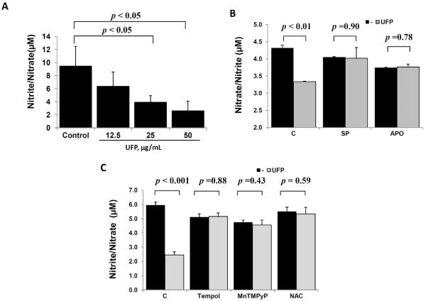
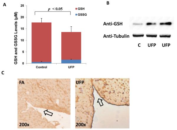
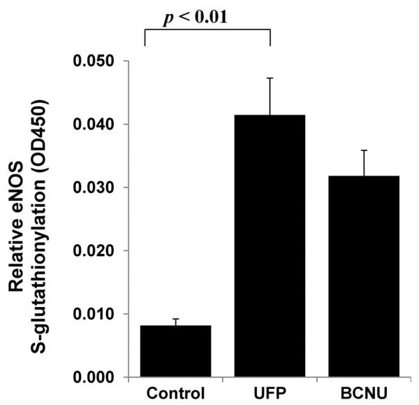
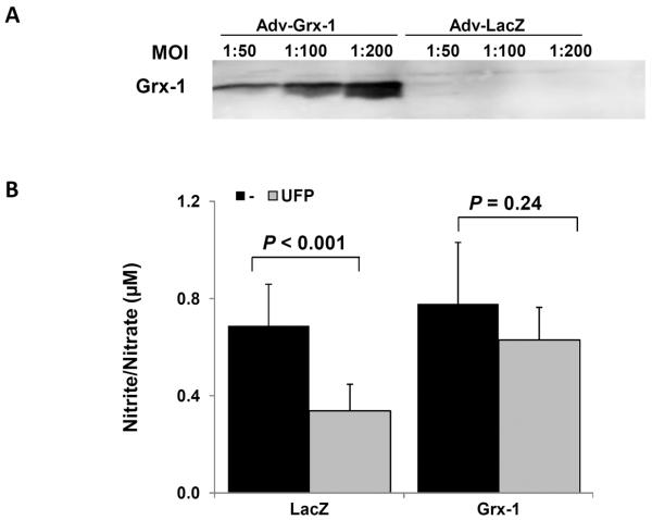
Exposure to airborne particulate pollutants is intimately linked to vascular oxidative stress and inflammatory responses with clinical relevance to atherosclerosis. Particulate matter (PM) has been reported to induce endothelial dysfunction and atherosclerosis. Here, we tested whether ambient ultrafine particles (UFP, diameter <200 nm) modulate eNOS activity in terms of nitric oxide (NO) production via protein S-glutathionylation. Treatment of human aortic endothelial cells (HAEC) with UFP significantly reduced NO production. UFP-mediated reduction in NO production was restored in the presence of JNK inhibitor (SP600125), NADPH oxidase inhibitor (Apocynin), anti-oxidant (N-acetyl cysteine), and superoxide dismutase mimetics (Tempol and MnTMPyP). UFP exposure increased the GSSG/GSH ratio and eNOS S-glutathionylation, whereas over-expression of Glutaredoxin-1 (to inhibit S-glutathionylation) restored UFP-mediated reduction in NO production by nearly 80%. Thus, our findings suggest that eNOS S-glutathionylation is a potential mechanism underlying ambient UFP-induced reduction of NO production.
Keywords: Air pollution; Endothelial dysfunction; Oxidative stress; S-glutathionylation; Ultrafine particles/UFP; eNOS.
Copyright © 2013 Elsevier Inc. All rights reserved.
Figures
Similar articles
-
Glutathionylation mediates angiotensin II-induced eNOS uncoupling, amplifying NADPH oxidase-dependent endothelial dysfunction.J Am Heart Assoc. 2014 Apr 22;3(2):e000731. doi: 10.1161/JAHA.113.000731. J Am Heart Assoc. 2014. PMID: 24755153 Free PMC article.
-
Nicorandil prevents endothelial dysfunction due to antioxidative effects via normalisation of NADPH oxidase and nitric oxide synthase in streptozotocin diabetic rats.Cardiovasc Diabetol. 2011 Nov 23;10:105. doi: 10.1186/1475-2840-10-105. Cardiovasc Diabetol. 2011. PMID: 22107602 Free PMC article.
-
Ultrafine particles from diesel engines induce vascular oxidative stress via JNK activation.Free Radic Biol Med. 2009 Mar 15;46(6):775-82. doi: 10.1016/j.freeradbiomed.2008.11.025. Epub 2008 Dec 11. Free Radic Biol Med. 2009. PMID: 19154785 Free PMC article.
-
Redox modulation of endothelial nitric oxide synthase by glutaredoxin-1 through reversible oxidative post-translational modification.Biochemistry. 2013 Sep 24;52(38):6712-23. doi: 10.1021/bi400404s. Epub 2013 Sep 11. Biochemistry. 2013. PMID: 23977830 Free PMC article.
-
Role of oxidative stress in the dysfunction of the placental endothelial nitric oxide synthase in preeclampsia.Redox Biol. 2021 Apr;40:101861. doi: 10.1016/j.redox.2021.101861. Epub 2021 Jan 19. Redox Biol. 2021. PMID: 33548859 Free PMC article. Review.
Cited by
-
A work group report on ultrafine particles (American Academy of Allergy, Asthma & Immunology): Why ambient ultrafine and engineered nanoparticles should receive special attention for possible adverse health outcomes in human subjects.J Allergy Clin Immunol. 2016 Aug;138(2):386-96. doi: 10.1016/j.jaci.2016.02.023. Epub 2016 Apr 6. J Allergy Clin Immunol. 2016. PMID: 27130856 Free PMC article. Review.
-
Xenobiotics Delivered by Electronic Nicotine Delivery Systems: Potential Cellular and Molecular Mechanisms on the Pathogenesis of Chronic Kidney Disease.Int J Mol Sci. 2022 Sep 7;23(18):10293. doi: 10.3390/ijms231810293. Int J Mol Sci. 2022. PMID: 36142207 Free PMC article. Review.
-
Oxidative stress and the cardiovascular effects of air pollution.Free Radic Biol Med. 2020 May 1;151:69-87. doi: 10.1016/j.freeradbiomed.2020.01.004. Epub 2020 Jan 7. Free Radic Biol Med. 2020. PMID: 31923583 Free PMC article. Review.
-
Accelerated Aging and Age-Related Diseases (CVD and Neurological) Due to Air Pollution and Traffic Noise Exposure.Int J Mol Sci. 2021 Feb 28;22(5):2419. doi: 10.3390/ijms22052419. Int J Mol Sci. 2021. PMID: 33670865 Free PMC article. Review.
-
The physiological roles of apolipoprotein J/clusterin in metabolic and cardiovascular diseases.Rev Endocr Metab Disord. 2014 Mar;15(1):45-53. doi: 10.1007/s11154-013-9275-3. Rev Endocr Metab Disord. 2014. PMID: 24097125 Review.
References
-
- Brooks MM, Chung SC, Helmy T, Hillegass WB, Escobedo J, Melsop KA, Massaro EM, McBane RD, Hyde P, Hlatky MA. Health Status After Treatment for Coronary Artery Disease and Type 2 Diabetes Mellitus in the Bypass Angioplasty Revascularization Investigation 2 Diabetes Trial. Circulation. 2010;122:1690–1699. - PMC - PubMed
-
- Hansen CS, Sheykhzade M, Moller P, Folkmann JK, Amtorp O, Jonassen T, Loft S. Diesel exhaust particles induce endothelial dysfunction in apoE−/− mice. Toxicol Appl Pharmacol. 2007;219:24–32. - PubMed
-
- Sun Q, Wang A, Jin X, Natanzon A, Duquaine D, Brook RD, Aguinaldo JG, Fayad ZA, Fuster V, Lippmann M, Chen LC, Rajagopalan S. Long-term air pollution exposure and acceleration of atherosclerosis and vascular inflammation in an animal model. JAMA. 2005;294:3003–3010. - PubMed
Publication types
MeSH terms
Substances
Grants and funding
LinkOut - more resources
Full Text Sources
Other Literature Sources
Research Materials
Miscellaneous
