Review
. 2013 Sep;12(4):982-95.
doi: 10.1016/j.arr.2013.05.008. Epub 2013 Jun 8.

Calcium dysregulation and neuroinflammation: discrete and integrated mechanisms for age-related synaptic dysfunction

Affiliations
Review

Calcium dysregulation and neuroinflammation: discrete and integrated mechanisms for age-related synaptic dysfunction

Diana M Sama et al. Ageing Res Rev. 2013 Sep.

Abstract

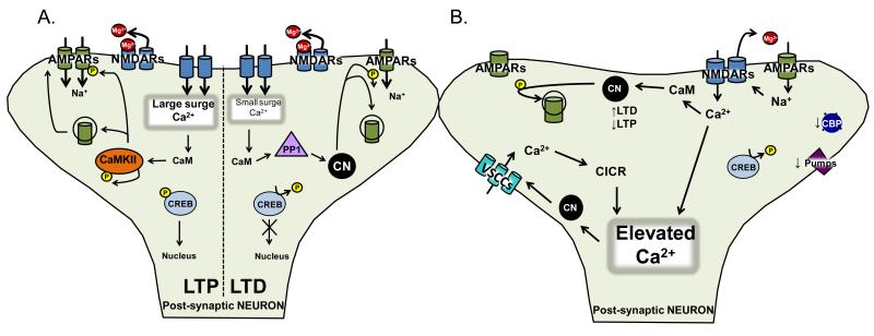
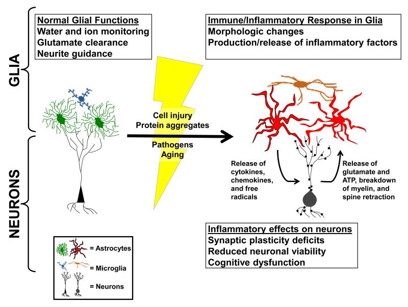
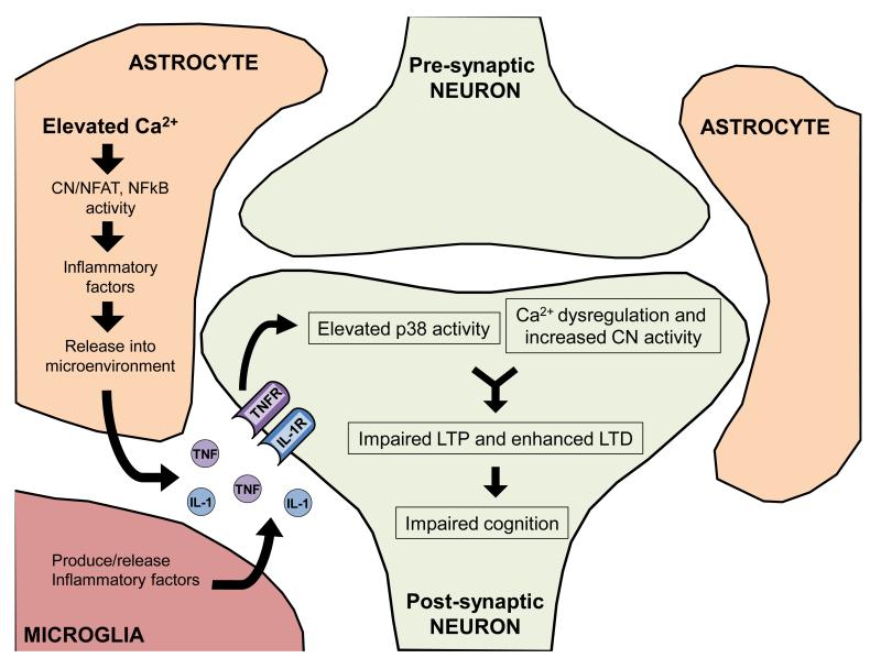
Some of the best biomarkers of age-related cognitive decline are closely linked to synaptic function and plasticity. This review highlights several age-related synaptic alterations as they relate to Ca(2+) dyshomeostasis, through elevation of intracellular Ca(2+), and neuroinflammation, through production of pro-inflammatory cytokines including interleukin-1 beta (IL-1β) and tumor necrosis factor-alpha (TNF-α). Though distinct in many ways, Ca(2+) and neuroinflammatory signaling mechanisms exhibit extensive cross-talk and bidirectional interactions. For instance, cytokine production in glial cells is strongly dependent on the Ca(2+) dependent protein phosphatase calcineurin, which shows elevated activity in animal models of aging and disease. In turn, pro-inflammatory cytokines, such as TNF, can augment the expression/activity of L-type voltage sensitive Ca(2+) channels in neurons, leading to Ca(2+) dysregulation, hyperactive calcineurin activity, and synaptic depression. Thus, in addition to discussing unique contributions of Ca(2+) dyshomeostasis and neuroinflammation, this review emphasizes how these processes interact to hasten age-related synaptic changes.

Keywords: Aging; Ca(2+); Cytokine; Neuroinflammation; Plasticity; Synapse.


Figures

Figure 1
Figure 1
A. Synaptic Plasticity. Long-term potentiation (LTP) and long-term depression (LTD) involve the activation of two glutamate receptors, α-amino-3-hydroxy-5-methylisoxazole-4-propionic acid receptors (AMPARs; green) and and N-methyl-D-aspartic acid receptors (NMDARs; blue). Both are permeable to Na+ and K+, while a subgroup of AMPARs and all NMDARs are permeable to Ca2+. Upon membrane depolarization by AMPARs, NMDARs expel a Mg2+-block (red), and permit Ca2+ entry into the postsynaptic cell. Both forms of plasticity require Ca2+ influx through NMDARs, though LTP generally requires a large surge in postsynaptic Ca2+, and LTD generally requires smaller Ca2+ influxes. As a result, the differing Ca2+ signals activate divergent biochemical cascades. Protein kinases, especially Ca2+/calmodulin-dependent protein kinase II (CaMKII; orange oval), have been strongly implicated in LTP, where they are involved in the phosphorylation (small yellow circle) and subsequent membrane insertion of glutamate receptors. Smaller elevations in Ca2+ are involved in LTD induction and activate protein phosphatases, namely Ca2+/calmodulin dependent protein phosphatase, calcineurin (CN; black circle), and/or protein phosphatase 1 (PP1; lavender triangle), which can be stimulated by CN activity. The balance in activity of protein kinases and phosphatases regulates the magnitude/duration of synaptic changes, and helps maintain the functional status of the synapse under basal conditions. B. Ca2+ dysregulation. Synaptic plasticity requires the activation of the appropriate biochemical cascades, and exquisite Ca2+ regulation. Under normal conditions, there are numerous mechanisms regulating intracellular Ca2+ concentrations. These include: voltage sensitive Ca2+ channels (VSCCs; aqua), a variety of receptors on the cell surface (NMDARs; light blue) or receptors on intracellular organelles which store Ca2+ (IP3Rs and ryanodine receptors), pumps (SERCA Ca2+; purple), binding proteins (calreticulin, BiP/grp78, grp94, and calnexin; royal blue), and intracellular organelles which store Ca2+ (endoplasmic reticulum and mitochondria). All of these mechanisms are impaired with aging, leading to increases in Ca2+ entry/release, and decreased Ca2+ expulsion/sequestration, leading to an elevation in Ca2+ concentration.
Figure 2
Figure 2. Neuroinflammation
Many astrocytes and microglia evidently morphologic changes from a “resting state” (green/blue) to an “activated state” (red/brown) under conditions of injury, disease, or aging (lightning bolt, yellow). When in a “resting state,” normal functions of glia include ion monitoring, glutamate clearance, and neurite guidance. Once astrocytes and microglia undergo a phenotype switch these functions are impaired and glia begin to produce and release immune/inflammatory mediators. The loss of glutamate transport and ion monitoring, together with the release of immune/inflammatory mediators, can lead to impaired synaptic plasticity, neuronal viability, and cognitive function.
Figure 3
Figure 3. The intersection of Ca2+ dysregulation and neuroinflammation
Ca2+ dysregulation in glia, especially astrocytes, can increase calcineurin (CN) activity and lead to the production and release of inflammatory factors. These factors can then initiate activation of the MAP kinase signaling pathways (p38) and Ca2+ dysregulation in neurons, leading to increased CN activity. Changes in neuronal Ca2+ signaling can further perpetuate Ca2+ dysregulation, synaptic plasticity deficits, and cognitive impairments.

References

    1. Abdul HM, Furman JL, Sama MA, Mathis DM, Norris CM. NFATs and Alzheimer’s Disease. Mol Cell Pharmacol. 2010;2:7–14. - PMC - PubMed
    1. Abdul HM, Sama MA, Furman JL, Mathis DM, Beckett TL, Weidner AM, Patel ES, Baig I, Murphy MP, LeVine H, 3rd, Kraner SD, Norris CM. Cognitive decline in Alzheimer’s disease is associated with selective changes in calcineurin/NFAT signaling. J Neurosci. 2009;29:12957–12969. - PMC - PubMed
    1. Ahn SH, Kim HJ, Jeong I, Hong YJ, Kim MJ, Rhie DJ, Jo YH, Hahn SJ, Yoon SH. Grape seed proanthocyanidin extract inhibits glutamate-induced cell death through inhibition of calcium signals and nitric oxide formation in cultured rat hippocampal neurons. BMC neuroscience. 2011;12:78. - PMC - PubMed
    1. Akiyama H, Barger S, Barnum S, Bradt B, Bauer J, Cole GM, Cooper NR, Eikelenboom P, Emmerling M, Fiebich BL, Finch CE, Frautschy S, Griffin WS, Hampel H, Hull M, Landreth G, Lue L, Mrak R, Mackenzie IR, McGeer PL, O’Banion MK, Pachter J, Pasinetti G, Plata-Salaman C, Rogers J, Rydel R, Shen Y, Streit W, Strohmeyer R, Tooyoma I, Van Muiswinkel FL, Veerhuis R, Walker D, Webster S, Wegrzyniak B, Wenk G, Wyss-Coray T. Inflammation and Alzheimer’s disease. Neurobiol Aging. 2000;21:383–421. - PMC - PubMed
    1. Albensi BC, Mattson MP. Evidence for the involvement of TNF and NF-kappaB in hippocampal synaptic plasticity. Synapse. 2000;35:151–159. - PubMed

Publication types