

Regulation of cell adhesion and migration by cell-derived matrices
- PMID: 23751565
- PMCID: PMC3780580
- DOI: 10.1016/j.yexcr.2013.05.030
Regulation of cell adhesion and migration by cell-derived matrices
Abstract
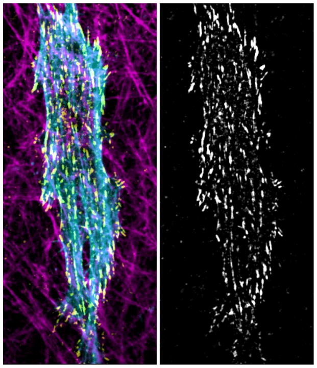
Three-dimensional in vitro extracellular matrix models provide a physiological alternative to regular two-dimensional cell culture, though they lack the full diversity of molecular composition and physical properties of whole-animal systems. Cell-derived matrices are extracellular matrices that are the product of matrix secretion and assembly by cells cultured at high density in vitro. After the removal of the cells that produced the matrix, an assembled matrix scaffold is left that closely mimics native stromal fiber organization and molecular content. Cell-derived matrices have been shown to impart in vivo-like responses to cells cultured in these matrices. In this review, we focus on mechanisms through which the distinct molecular and topographical composition of cell-derived matrices directs cellular behavior, specifically through regulation of cell-matrix adhesions and subsequent contributions to the process of cell migration.
Keywords: 3D cell migration; Cell-derived matrix (CDM); Extracellular matrix; Fibrillar topography; Matrix adhesions; Matrix elasticity.
Published by Elsevier Inc.
Figures
Similar articles
-
Direct comparisons of the morphology, migration, cell adhesions, and actin cytoskeleton of fibroblasts in four different three-dimensional extracellular matrices.Tissue Eng Part A. 2011 Mar;17(5-6):713-24. doi: 10.1089/ten.TEA.2010.0273. Epub 2010 Dec 7. Tissue Eng Part A. 2011. PMID: 20929283 Free PMC article.
-
Podosome-type adhesions and focal adhesions, so alike yet so different.Eur J Cell Biol. 2008 Sep;87(8-9):491-506. doi: 10.1016/j.ejcb.2008.02.012. Epub 2008 Apr 15. Eur J Cell Biol. 2008. PMID: 18417250 Review.
-
Micro-environmental control of cell migration--myosin IIA is required for efficient migration in fibrillar environments through control of cell adhesion dynamics.J Cell Sci. 2012 May 1;125(Pt 9):2244-56. doi: 10.1242/jcs.098806. Epub 2012 Feb 10. J Cell Sci. 2012. PMID: 22328520 Free PMC article.
-
Direct comparison of five different 3D extracellular matrix model systems for characterization of cancer cell migration.Cancer Rep (Hoboken). 2020 Oct;3(5):e1257. doi: 10.1002/cnr2.1257. Epub 2020 Jun 8. Cancer Rep (Hoboken). 2020. PMID: 33085847 Free PMC article.
-
Dimensions and dynamics in integrin function.Braz J Med Biol Res. 2003 Aug;36(8):959-66. doi: 10.1590/s0100-879x2003000800001. Epub 2003 Jul 23. Braz J Med Biol Res. 2003. PMID: 12886449 Review.
Cited by
-
Stimuli-Responsive Hydrogels: The Dynamic Smart Biomaterials of Tomorrow.Macromolecules. 2023 Oct 18;56(21):8377-8392. doi: 10.1021/acs.macromol.3c00967. eCollection 2023 Nov 14. Macromolecules. 2023. PMID: 38024154 Free PMC article. Review.
-
Techniques for assessing 3-D cell-matrix mechanical interactions in vitro and in vivo.Exp Cell Res. 2013 Oct 1;319(16):2470-80. doi: 10.1016/j.yexcr.2013.06.018. Epub 2013 Jun 29. Exp Cell Res. 2013. PMID: 23819988 Free PMC article. Review.
-
Comparative studies of cellular viability levels on 2D and 3D in vitro culture matrices.Cytotechnology. 2018 Feb;70(1):261-273. doi: 10.1007/s10616-017-0139-7. Epub 2017 Sep 18. Cytotechnology. 2018. PMID: 28924965 Free PMC article.
-
Generation of 3D Collagen Gels with Controlled Diverse Architectures.Curr Protoc Cell Biol. 2016 Sep 1;72:10.20.1-10.20.16. doi: 10.1002/cpcb.9. Curr Protoc Cell Biol. 2016. PMID: 27580704 Free PMC article.
-
Cell-derived extracellular matrix-coated silk fibroin scaffold for cardiogenesis of brown adipose stem cells through modulation of TGF-β pathway.Regen Biomater. 2020 Aug;7(4):403-412. doi: 10.1093/rb/rbaa011. Epub 2020 Apr 24. Regen Biomater. 2020. PMID: 32793385 Free PMC article.
References
-
- Yamada KM, Cukierman E. Modeling tissue morphogenesis and cancer in 3D. Cell. 2007;130:601–610. - PubMed
-
- Soucy PA, Romer LH. Endothelial cell adhesion, signaling, and morphogenesis in fibroblast-derived matrix. Matrix Biol. 2009;28:273–283. - PubMed
-
- Zwolinski CM, Ellison KS, Depaola N, Thompson DM. Generation of cell-derived three dimensional extracellular matrix substrates from two dimensional endothelial cell cultures. Tissue Eng Part C Methods. 2011;17:589–595. - PubMed
Publication types
MeSH terms
Grants and funding
LinkOut - more resources
Full Text Sources
Other Literature Sources
Miscellaneous
